Integration of PET/CT Radiomics and Semantic Features for Differentiation between Active Pulmonary Tuberculosis and Lung
- PDF / 2,984,940 Bytes
- 12 Pages / 595.276 x 790.866 pts Page_size
- 90 Downloads / 304 Views
RESEARCH ARTICLE
Integration of PET/CT Radiomics and Semantic Features for Differentiation between Active Pulmonary Tuberculosis and Lung Cancer Dongyang Du,1 Jiamei Gu,2 Xiaohui Chen,2 Wenbing Lv,1 Qianjin Feng,1 Arman Rahmim,3,4 Hubing Wu,2 Lijun Lu 1 1
School of Biomedical Engineering and Guangdong Provincial Key Laboratory of Medical Image Processing, Southern Medical University, Guangzhou, 510515, Guangdong, China 2 Nanfang PET Center, Nanfang Hospital, Southern Medical University, Guangzhou, 510515, Guangdong, China 3 Departments of Radiology and Physics, University of British Columbia, Vancouver, BC, V6T 1Z1, Canada 4 Department of Integrative Oncology, BC Cancer Research Centre, Vancouver, BC, V5Z 1L3, Canada
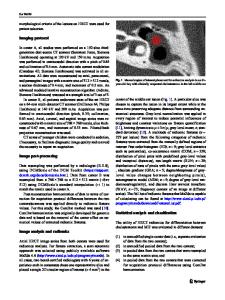
Abstract Purpose: We aim to accurately differentiate between active pulmonary tuberculosis (TB) and lung cancer (LC) based on radiomics and semantic features as extracted from pre-treatment positron emission tomography/X-ray computed tomography (PET/CT) images. Procedures: A total of 174 patients (77/97 pulmonary TB/LC as confirmed by pathology) were retrospectively selected, with 122 in the training cohort and 52 in the validation cohort. Four hundred eighty-seven radiomics features were initially extracted to quantify phenotypic characteristics of the lesion region in both PET and CT images. Eleven semantic features were additionally defined by two experienced nuclear medicine physicians. Feature selection was performed in 5 steps to enable derivation of robust and effective signatures. Multivariable logistic regression analysis was subsequently used to develop a radiomics nomogram. The calibration, discrimination, and clinical usefulness of the nomogram were evaluated in both the training and independent validation cohorts. Results: The individualized radiomics nomogram, which combined PET/CT radiomics signature with semantic features, demonstrated good calibration and significantly improved the diagnostic performance with respect to the semantic model alone or PET/CT signature alone in training cohort (AUC 0.97 vs. 0.94 or 0.91, p = 0.0392 or 0.0056), whereas did not significantly improve the performance in validation cohort (AUC 0.93 vs. 0.89 or 0.91, p = 0.3098 or 0.3323). Conclusion: The radiomics nomogram showed potential for individualized differential diagnosis between solid active pulmonary TB and solid LC, although the improvement of performance was not significant relative to semantic model. Key words: Radiomics, FDG-PET/CT, Active pulmonary tuberculosis, Lung cancer, Diagnosis
Introduction Electronic supplementary material The online version of this article (https:// doi.org/10.1007/s11307-020-01550-4) contains supplementary material, which is available to authorized users. Correspondence to: Hubing Wu; e-mail: [email protected], Lijun Lu; email: [email protected]
Tuberculosis (TB) is a global public health threat, is prevalent in numerous developing countries, and is the second-most common cause of death from infectious disease [1, 2]. Pulmonary TB, as a kind of communicable disease,
Data Loading...