MRI-Based radiomics nomogram for differentiation of benign and malignant lesions of the parotid gland
- PDF / 2,210,438 Bytes
- 11 Pages / 595.276 x 790.866 pts Page_size
- 97 Downloads / 392 Views
HEAD AND NECK
MRI-Based radiomics nomogram for differentiation of benign and malignant lesions of the parotid gland Ying-mei Zheng 1 & Jian Li 2 & Song Liu 3 & Jiu-fa Cui 3 & Jin-feng Zhan 3 & Jing Pang 3 & Rui-zhi Zhou 3 & Xiao-li Li 3 & Cheng Dong 3 Received: 13 July 2020 / Revised: 31 August 2020 / Accepted: 5 November 2020 # European Society of Radiology 2020
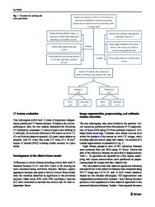
Abstract Objectives Preoperative differentiation between benign parotid gland tumors (BPGT) and malignant parotid gland tumors (MPGT) is important for treatment decisions. The purpose of this study was to develop and validate an MRI-based radiomics nomogram for the preoperative differentiation of BPGT from MPGT. Methods A total of 115 patients (80 in training set and 35 in external validation set) with BPGT (n = 60) or MPGT (n = 55) were enrolled. Radiomics features were extracted from T1-weighted and fat-saturated T2-weighted images. A radiomics signature model and a radiomics score (Rad-score) were constructed and calculated. A clinical-factors model was built based on demographics and MRI findings. A radiomics nomogram model combining the Rad-score and independent clinical factors was constructed using multivariate logistic regression analysis. The diagnostic performance of the three models was evaluated and validated using ROC curves on the training and validation datasets. Results Seventeen features from MR images were used to build the radiomics signature. The radiomics nomogram incorporating the clinical factors and radiomics signature had an AUC value of 0.952 in the training set and 0.938 in the validation set. Decision curve analysis showed that the nomogram outperformed the clinical-factors model in terms of clinical usefulness. Conclusions The above-described radiomics nomogram performed well for differentiating BPGT from MPGT, and may help in the clinical decision-making process. Key Points • Differential diagnosis between BPGT and MPGT is rather difficult by conventional imaging modalities. • A radiomics nomogram integrated with the radiomics signature, clinical data, and MRI features facilitates differentiation of BPGT from MPGT with improved diagnostic efficacy. Keywords Parotid neoplasms . Magnetic resonance imaging . Radiomics
Ying-mei Zheng and Jian Li contributed equally to this work. Ying-mei Zheng and Jian Li are collaborative first author * Cheng Dong [email protected] 1
Health Management Center, The Affiliated Hospital of Qingdao University, No.16, Jiangsu Road, Qingdao 266000, China
2
Department of Radiology, The University of Hong Kong - Shenzhen Hospital, No.1, Haiyuan Road, Futian District, Shenzhen 518000, China
3
Department of Radiology, The Affiliated Hospital of Qingdao University, No.16, Jiangsu Road, Qingdao 266000, China
Abbreviations 3-D ANOVA BPGT CI DCA DLI FNA fs-T2WI GLCM GLDM GLRLM GLSZM ICC IST
Three-dimensional Analysis of variance Benign parotid gland tumors Confidence interval Decision curve analysis Deep lobe involved Fine needle aspiration Fat-saturated T2-weighted images Gray-level co-occurrence matr
Data Loading...