Ionophore properties of valinomycin in the model bilayer lipid membrane 1. Selectivity towards a cation
- PDF / 1,632,542 Bytes
- 10 Pages / 595.276 x 790.866 pts Page_size
- 27 Downloads / 355 Views
ORIGINAL PAPER
Ionophore properties of valinomycin in the model bilayer lipid membrane 1. Selectivity towards a cation ZhangFei Su 1 & Dusan Mrdenovic 1,2 & Slawomir Sek 1,3 & Jacek Lipkowski 1 Received: 6 June 2020 / Revised: 18 July 2020 / Accepted: 19 July 2020 # Springer-Verlag GmbH Germany, part of Springer Nature 2020
Abstract The electrochemical impedance spectroscopy (EIS) and polarization-modulation infrared reflection absorption spectroscopy (PM-IRRAS) techniques were employed to study the ionophore properties of valinomycin in a model floating bilayer lipid membrane (fBLM) in perchlorate supporting electrolytes with potassium and sodium cations. Valinomycin decreases the membrane resistance of the fBLM in KClO4 solution by about 180 times, while it has a negligible effect on the membrane resistivity in NaClO4 solution. The IR spectra indicate that valinomycin forms a complex with K+, but not with Na+. The valinomycin-K+ complex adopts a small tilt angle with respect to the electrode surface normal and is well interdigitated between the acyl chains of the bilayer. The EIS and PM-IRRAS results indicate that valinomycin forms complexes with K+ and transports K+ across the lipid bilayer. This transport is potential independent and hence has a passive character. Keywords Valinomycin . Floating bilayer lipid membrane . Cation . EIS . PM-IRRAS
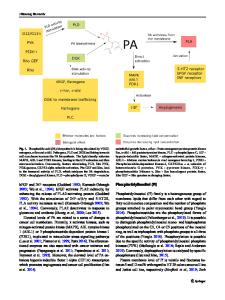
Introduction Ionophores are lipid-soluble molecules, which can bind and transport ions across natural and artificial membranes [1]. Valinomycin is an antibiotic cyclic neutral peptide, which acts as a carrier-type ionophore in cell membranes by selectively transporting K+ across the membrane [2–4]. Valinomycin consists of twelve alternating amino acids, whose structure is shown in Scheme 1. Valinomycin has high cation selectivity
Dedicated to Professor Fritz Scholz on the occasion of his 65th birthday and in recognition of his contributions to electrochemistry Electronic supplementary material The online version of this article (https://doi.org/10.1007/s10008-020-04777-x) contains supplementary material, which is available to authorized users. * Jacek Lipkowski [email protected] 1
Department of Chemistry, University of Guelph, Guelph, Ontario N1G 2W1, Canada
2
Institute of Physical Chemistry, Polish Academy of Sciences, Kasprzaka 44/52, 01-224 Warsaw, Poland
3
Faculty of Chemistry, Biological and Chemical Research Centre, University of Warsaw, Żwirki i Wigury 101, 02-089 Warsaw, Poland
by forming a complex with alkali metal cations, such as K+, Rb+, and Cs+, but not Na+ or Li+ [5]. The valinomycin-K+ complex forms a bracelet conformation, in which K+ stays in the center of the valinomycin ring cavity and binds to six ester C=O groups [6]. In contrast, free (noncomplex) valinomycin adopts a propeller conformation [7]. Infrared spectroscopy is a powerful tool to investigate the structure of valinomycin in different environments and with different cations. In phospholipid bilayers, the IR spectrum of free valinomycin displays two amide I bands and two ester C=O
Data Loading...