Is Decompressive Craniectomy a Risk Factor for Ventriculomegaly?
Objective: Decompressive craniectomy (DC) is an established therapeutic option following severe traumatic brain injury (TBI). However, several delayed complications of DC have been reported, including ventriculomegaly, which can lead to poor patient outco
- PDF / 234,866 Bytes
- 3 Pages / 595.28 x 790.87 pts Page_size
- 21 Downloads / 298 Views
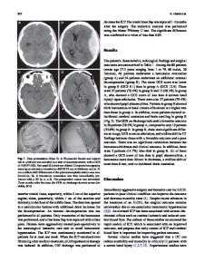
Abstract Objective: Decompressive craniectomy (DC) is an established therapeutic option following severe traumatic brain injury (TBI). However, several delayed complications of DC have been reported, including ventriculomegaly, which can lead to poor patient outcomes. Nevertheless, ventriculomegaly can occur after TBI even without DC. The aim of the present study was to investigate the influence of DC on ventriculomegaly. Material and Methods: Adult male Sprague–Dawley rats (300–400 g) were subjected to lateral fluid percussion injury using a fluid percussion device. Rats were randomly divided into four groups: sham, craniectomized without trauma (D group), traumatized without DC (FPI group), and craniectomized immediately after trauma (FPI + D group). On day 28 of recovery, ventricular volumes were measured by image analysis. Results: There was no significant difference in ventricular size between the sham group and the D group animals or between the FPI group and the FPI + D group animals. Conclusion: These data suggest that DC may not be a risk factor for ventriculomegaly after TBI. Keywords Ventriculomegaly • Decompressive craniectomy • Traumatic brain injury • Rat
Introduction Decompressive craniectomy (DC) is an established therapeutic option following severe traumatic brain injury (TBI), and is widely used in the treatment of malignant post-
S. Takeuchi (*), K. Nagatani, K. Wada, H. Nawashiro, N. Otani, H. Osada, H. Kobayashi, T. Suzuki, and K. Shima Department of Neurosurgery, National Defense Medical College, 3-2 Namiki, Tokorozawa, Saitama 359-8513, Japan e-mail: [email protected]
traumatic brain edema [1, 12, 14, 16]. The beneficial effects of DC in reducing intracranial pressure (ICP) are well documented, and DC can also contribute to improvements in cerebral compliance, cerebral oxygen supply, cerebral blood perfusion, and various abnormal computed tomography signs [1, 12, 14, 16]. However, several delayed complications of DC have been reported [2, 4, 5, 7, 15, 20], including ventriculomegaly, which can lead to a poor outcome for the patients [2, 4, 5]. On the other hand, ventriculomegaly can occur after TBI even without DC [3, 6, 9, 13], and clinical studies failed to examine whether DC itself could aggravate ventriculomegaly. The aim of the present study was to investigate the influence of DC on ventriculomegaly.
Materials and Methods Animals and Experimental Procedures All experimental procedures were approved by the Animal Care and Use Committee of the National Defense Medical College. Sprague–Dawley (SD) rats (male, 320–390 g; 9–10 weeks of age) were used. The rats were housed in individual cages under controlled environmental conditions (12/12 h light/dark cycle, 20–22 °C; room temperature) with food and water freely available, for 1 week before the experimental surgery. The rats were anesthetized with isofluorane (1.5 %) in a 30 % oxygen to 70 % nitrous oxide gas mixture via a nose cone and were fixed in a stereotaxic frame for the procedure. A 4.8 mm craniotomy was made over the rig
Data Loading...