Low Voltage Table-Top Electron Microscopy of Polymer and Organic Molecular Thin Films
- PDF / 699,922 Bytes
- 6 Pages / 612 x 792 pts (letter) Page_size
- 52 Downloads / 270 Views
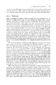
Low Voltage Table-Top Electron Microscopy of Polymer and Organic Molecular Thin Films David C. Martin, Lawrence F. Drummy, Junyan Yang, and Eva Coufalova* The Macromolecular Science and Engineering Center and the Departments of Materials Science and Engineering and Biomedical Engineering The University of Michigan, 2022 H. H. Dow Building, Ann Arbor, MI 48109-2136 *Delong Instruments s.r.o., Brno, Czech Republic INTRODUCTION There is intense current interest in the use of polymer and organic molecular materials for high technology devices including transistors, biosensors, and actuators [1]. In order for these materials to achieve their ultimate potential it is imperative to obtain detailed information about their microstructure, especially in the thin film forms central to technological applications. Our research group has been actively developing techniques for high-resolution electron optical examinations of polymers and organic materials, with particular emphasis on low dose highresolution electron microscopy (HREM) [2]. While this method of examining organic materials’ structure has proven to be particularly powerful, current generations of electron microscopes suffer from several problems. They are expensive, require considerable amounts of space, and are time-consuming and fairly difficult to operate. Furthermore, the high voltages normally used in conventional electron microscopes (200–400 kV) provide little contrast when imaging thin films composed of low atomic number components. Recently, Delong Instruments in Brno, Czech Republic (www.dicomps.com) has developed a new electron microscope that overcomes many of these disadvantages [3]. Their design consists of a table-top sized, low voltage (~5 kV) electron microscope (LVEM) capable of operating in transmission electron microscopy (TEM), scanning transmission electron microscopy (STEM), scanning electron microscopy (SEM), and electron diffraction (ED) modes. The instrument is constructed with a small Schottky field-emission gun, permanent magnet condenser and objective lenses, electrostatic projector lenses, and a YAG screen for image acquisition using conventional optics and a CCD camera. The small footprint of the device Figure 1. The low voltage electron microscope developed by Delong makes it attractive for Instruments. The nominal operating voltage is ~5 kV. Shown on the situations where space is at screen are experimental images of polyethylene single crystals a premium. The small showing their well-known lamellar morphology. FF6.4.1
2
Critical dose (C/cm )
volume of the vacuum chamber makes it possible to maintain an excellent vacuum (10-8 torr at the FEG and 10-7 near the sample) and provides for rapid sample exchange times. The pumping system includes two small ion pumps connected to the optical chamber, with a turbomolecular pump on the floor behind the unit. Although there is a slight increase in the electron wavelength at these lower voltages (0.017 nm at 5 kV vs. 0.0016 nm at 400 kV), there is still more than sufficient instrumental resolu
Data Loading...