Magnetron Sputtering Deposition of Calcium Phosphate Films with Nanoscale Grain Morphology in their Surface
- PDF / 441,969 Bytes
- 6 Pages / 612 x 792 pts (letter) Page_size
- 23 Downloads / 349 Views
0975-DD06-08
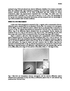
Magnetron Sputtering Deposition of Calcium Phosphate Films with Nanoscale Grain Morphology in their Surface Wilfredo OtaÒo1, VÌctor M. Pantojas1, Juan M. Figueroa1, Darimar Hern·ndez1, and Alejandro RodrÌguez-Navarro2 1 Mathematics-Physics, University of Puerto Rico at Cayey, 205 A. Barcelo Avenue, Cayey, 00736, Puerto Rico 2 Universidad de Granada, Instituto Andaluz de Ciencias de la Tierra-CSIC, Campus de Fuentenueva, Granada, 18002, Spain ABSTRACT Hydroxyapatite (HA) is a calcium phosphate mineral analogous to the mineral part of bone. This similarity makes this material bioactive and suitable to coat medical implants. However, at present, there is not a coating technique which gives the coated implant the desired properties and long life required for medical implants. In an effort to produce HA coatings with improved properties, calcium phosphate films were prepared using magnetron sputtering deposition on a silicon substrate at 600∞C. Initial efforts resulted in the deposition of amorphous films with a distinctive grain-like surface morphology. The morphological grain size was studied using SEM, and it was found that the average diameter value of the round shaped grains could be controlled by adjusting the deposition time. Increasing the deposition time increases the mean grain diameter. EDS spectra showed the unintentional addition of carbon, iron and nickel to the samples during deposition. After eliminating the impurities, it was possible to prepare calcium phosphate films in the HA phase but without the grain-like surface morphology. These results suggested that the impurities prevented the formation of the calcium phosphate HA phase while acting as nuclei for the heterogeneous nucleation of the grains. This is an important result where the deposition process parameters can be controlled to functionalize the films in order to produce distinctive nanoscale features in the surface morphology.
INTRODUCTION The calcium phosphate crystalline phase hydroxyapatite (HA) is a bioactive material with important biomedical applications. The most important ones are its use as a bone graft substitute in dental and orthopedic applications and as a coating of biomedical implants [1]. HA coatings have been prepared using different techniques such as plasma spray [2-4], laser ablation [5,6], dual ion beam [7,8], and sputtering deposition [9-15], among others, with different degrees of success. Plasma spray is the technique actually used commercially to coat medical implants with HA and, consequently, those coatings are the most studied ones. Nevertheless, the latter technique produces coatings with poor mechanical properties and weak adhesion to the substrate interface, resulting in delamination and debris formation [16].
Sputtering based techniques are widely used in many industries (e.g., to coat cutting tools). The coatings produced by sputtering are very dense, and have excellent adhesion to the substrate and superior mechanical properties as well as a long life. The use of sputtering techniques is a goo
Data Loading...