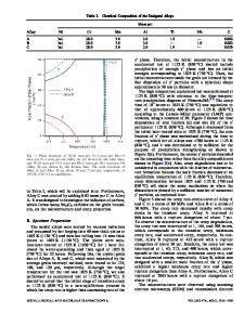
Measuring creep damage using microradiography
- PDF / 3,918,155 Bytes
- 9 Pages / 612 x 828 pts Page_size
- 82 Downloads / 390 Views
I.
INTRODUCTION
PREVIOUS investigations of creep damage have utilized techniques such as optical microscopy, scanning electron microscopy (SEM), and dilatometry. While each of these techniques has advantages, each also has certain limitations. Optical microscopy and SEM can both be used to examine fracture surfaces and polished sections for creep damage. While the resolution of these techniques, particularly using SEM, is very good, the results may not be representative of the bulk material since only a surface is examined. Additionally, artifacts may be introduced into a sample when preparing polished sections. Dilatometry can be used to determine the amount of creep damage in a bulk sample; however, it gives no information on the size and spatial distribution of the damage. A technique which can be applied to a bulk sample and which can also give information on the size and distribution of creep damage would be an excellent complement to existing techniques. Microradiography is such a technique. When a sample is placed in an X-ray beam, the directly transmitted beam contains the radiographic image of the sample, in which intensity variations are formed by differential absorption of the X-ray beam by different regions of the sample. Surface roughness, second phase particles and other regions of different composition, variations in sample thickness, and variations in density will all lead to differential absorption which causes intensity variations in a radiographic image. Microradiography refers to the investigation of fine structure on these radiographs, generally using a light microscope. Microradiography was first employed around 1900 to study biological specimens ~'2 and some alloys. 3 Since then microradiography has had limited use in the field of materials science due to the geometric resolution limitations and the long exposure times required when using conventional X-ray sources. With the increased availability of high intensity synchrotron radiation, interest in microradi0graphy has been renewed. The X-ray intensity available from a synchrotron source is over three orders of magni-
J.E. BENCI, Graduate Research Fellow, and D.P. POPE, Professor, are with the Department of Materials Science and Engineering, The University of Pennsylvania, 3231 Walnut Street, Philadelphia, PA 19104. This paper is based on a presentation made in the symposium "Crack Propagation under Creep and Creep-Fatigue" presented at the TMS/AIME fall meeting in Orlando, FL, in October 1986, under the auspices of the ASM Flow and Fracture Committee. METALLURGICALTRANSACTIONS A
tude greater than that available from a conventional X-ray source. Purushothaman et al. 4 performed preliminary experiments to test the feasibility of using synchrotron microradiography to study internal damage in metals. They concluded that much more damage can be detected using microradiography than is visible on the surface of the samples using microscopy. The purpose of the study reported here is to determine what new information about creep damage can be reve
Data Loading...