Microscopic Characterizations of Nanostructured Silicon Thin Films for Solar Cells
- PDF / 497,904 Bytes
- 9 Pages / 432 x 648 pts Page_size
- 49 Downloads / 398 Views
Microscopic Characterizations of Nanostructured Silicon Thin Films for Solar Cells Antonín Fejfar1, Petr Klapetek2, Jakub Zlámal3, Aliaksei Vetushka1, Martin Ledinský1 and Jan Kočka1 1
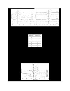
Institute of Physics of the Academy of Sciences of the Czech Republic, v.v.i, Cukrovarnická 10, 162 00 Prague 6, Czech Republic 2 Czech Metrology Institute, Okružní 31, 638 00 Brno, Czech Republic 2 Brno University of Technology, Technická 2, 616 69 Brno, Czech Republic ABSTRACT Microscopic characterization of mixed phase silicon thin films by conductive atomic force microscopy (C-AFM) was used to study the structure composed of conical microcrystalline grains dispersed in amorphous matrix. C-AFM experiments were interpreted using simulations of electric field and current distributions. Density of absorbed optical power was calculated by numerically solving the Maxwell equations. The goal of this study is to combine both models in order to simulate local photoconductivity for understanding the charge photogeneration and collection in nanostructured solar cells. INTRODUCTION Individual grains in silicon thin films prepared close to the border between the amorphous and microcrystalline growth have sizes from 10 to ~1000 nm or more. The grains may be aggregated in typical cones connected to each other via boundaries or surrounded by amorphous tissue. The cones end on the surface by nearly spherical caps [1,2] with a cauliflower structure, as shown in Fig. 1. U J
d a-Si
side view:
r
d incubation
top view: a-Si:H
bisectors grain boundaries
c-Si
nucleus
nearest neighbor (Delaunay) triangulation
Figure 1 Left: Scheme of the typical structure of mixed phase Si layer. Right: Scanning electron microscopy image showing an example of mixed phase microcrystalline silicon layer. Spherical caps of the microcrystalline Si grains with a cauliflower structure can be seen together with surrounding columnar amorphous phase [3].
313
Macroscopic properties and thus also the operation of the devices based on nanostructural silicon are determined by the properties of structural components, their spatial arrangement and mutual interaction. Large differences in conductivities of the components lead to substantial redistribution of internal electrical fields [4]. Local variation of the internal fields are usually not taken into account for discussing the operation of thin film solar cells, in spite of the fact that both the thickness and spatial features are comparable to photon wavelengths, leading to pronounced near field effects [5]. Since more than 10 years we have used the tip of atomic force microscopy (AFM) cantilever in contact mode as a local contact (i.e., Conductive AFM or C-AFM [6]) to measure the structure and local electronic properties with spatial resolution down to several nanometers [7,8]. In the course of the study we discovered the differences between the conductive AFM measurements in UHV and ambient atmosphere [3] and clarified how the applied bias may change the surface properties of the samples, even leading to fundamental
Data Loading...