Microstructural studies of laser irradiated graphite surfaces a)
- PDF / 1,862,939 Bytes
- 9 Pages / 593.28 x 841.68 pts Page_size
- 102 Downloads / 356 Views
J. Steinbeckb) Department of Physics, Massachusetts Institute of Technology, Cambridge, Massachusetts 02139
M. S. Dresselhaus Department of Physics and Department of Electrical Engineering and Computer Science, Massachusetts Institute of Technology, Cambridge, Massachusetts 02139 (Received 30 October 1989; accepted 2 February 1990)
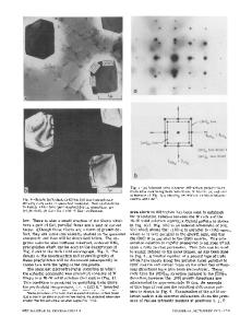
The structure of pulsed laser irradiated graphite surfaces has been elucidated. The pulse fluences range up to 4 J cm"2 with durations no longer than 30 ns. The region exterior to the irradiated spot is littered with —0.1 jam diameter carbon spheroids. The boundary region is characterized by both spheroids and torn layers 1-5 fim in lateral extent. The central region displays carbon spheroids and surface upheavals. The carbon spheroids are attributed to hydrodynamic sputtering of carbon. The surface upheavals and torn carbon layers are attributed to constrained thermal expansion and contraction of the irradiated region. It is estimated that a nearly instantaneous 1000 °C temperature change is necessary to cause the observed surface deformation. Pulse fluences in excess of 0.8 J cm"2 cause a thin layer of carbon to melt. This is proven by the fact that the irradiated layer in the solid phase has a turbostratic structure. Electron diffraction experiments and dark-field imaging experiments show that the basal plane grain size of the resolidified material varies from —20 A at the melt threshold to —100 A for samples irradiated with 4.0 J cm"2.
I. INTRODUCTION
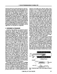
The high temperature phases and properties of carbon, the most refractory material known to man, have long been of interest. The nature of the high temperature-low pressure region of the carbon phase diagram is still known with little certainty.1 The high melting temperature of carbon (estimated to be —4500 K2) and the high vapor pressure near the melting point prove to be substantial experimental barriers to effectively probing this region of the P-T phase diagram. It is difficult to confine a —4500 K melt pool of carbon (what could be used for a crucible?). Hence most direct experimental work on the structure and properties of liquid carbon have involved rapid heating of carbon either by electrical currents3'4 or by short intense laser pulses.5"7 Previous work by Braunstein et al. on the problem using Rutherford backscattering spectrometry (RBS), Raman spectroscopy, and ion channeling has led to values of the disordered layer thickness as a function of laser pulse fluence,8 as shown in Fig. 1. These investigators concluded that liquid carbon was formed when a)Portions
of the text and results were presented at the 1985 and 1987 Fall Meetings of the Materials Research Society. b)Current address: Rome Air Development Center, Hanscom AFB, Massachusetts 01731. 980
http://journals.cambridge.org
J. Mater. Res., Vol. 5, No. 5, May 1990
Downloaded: 14 Mar 2015
samples were irradiated by a 30 ns pulsed ruby laser (A = 6943 A) with fluences greater than 0.6 J cm"2. Further evidence for melting can be obtained by direct microscopic exami
Data Loading...