Multiple cardiac complications associated with collagen disease
- PDF / 595,869 Bytes
- 2 Pages / 595.276 x 790.866 pts Page_size
- 96 Downloads / 341 Views
CASE IMAGE IN CARDIOVASCULAR ULTRASOUND
Multiple cardiac complications associated with collagen disease Makiko Suzuki1 · Hidekazu Tanaka1 · Shun Yokota1 · Tetsuya Hara1 · Yo Ueda2 · Ken‑ichi Hirata1 Received: 16 July 2020 / Revised: 21 October 2020 / Accepted: 4 November 2020 © Japanese Society of Echocardiography 2020
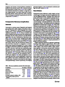
A 35-year-old woman under treatment of collagen disease due to scleroderma was admitted to the Division of Rheumatology and Clinical Immunology in our hospital because of slight fever, arthralgia, and multiple lymphadenopathy. Transthoracic echocardiography was performed on the 3rd hospital day because of the development of dyspnea, because the enlargement of the heart in a chest X-ray, and elevated creatine kinase (2,250 IU/L) and C-reactive protein (19.3 mg/dL). It revealed a Takotsubo cardiomyopathy-like left ventricular (LV) wall motion abnormality with an LV ejection fraction (LVEF) as low as 31% (Fig. 1a). None/ trivial mitral regurgitation (MR), aortic regurgitation (AR), and tricuspid regurgitation (TR) were shown (Fig. 1b). However, previous transthoracic echocardiography showed no specific abnormality with an LVEF as good as 68% (Fig. 1a). Coronary angiography did not show significant stenosis, and pulmonary capillary wedge pressure, mean pulmonary artery pressure, and cardiac index were 11 mmHg, 17 mmHg, and 2.82 L/min/m2, respectively. Moreover, the histological findings of hematoxylin–eosin staining obtained from the right ventricular apex showed mild myocardial fibrosis. Cardiac magnetic resonance imaging also showed a Takotsubo cardiomyopathy-like LV wall motion abnormality. Furthermore, patchy late gadolinium enhancement was observed in the endocardium of basal-mid lateral and the apex, suggesting the endomyocardial fibrosis due to collagen disease rather than virus myocarditis or Takotsubo cardiomyopathy. The patient was then diagnosed as acute exacerbation of existing scleroderma and further complications of systemic lupus erythematosus, Sjogren’s syndrome, and antiphospholipid antibody syndrome by attending doctors.
The patient received a 3-day course of intravenous methylprednisolone (1000 mg daily) followed by a declining, maintenance dose of 12.5 mg prednisolone daily, and immunosuppressive therapy consisting of mycophenolate mofetil at a dose of 1000 mg daily. Transthoracic-echocardiography was re-examined on the 124th hospital day and showed that a Takotsubo cardiomyopathy-like LV wall motion abnormality was similar with LVEF of 29%, and MR, AR and TR were worsened (Fig. 1c). Interestingly, the calcification of the posterior papillary muscle was newly appeared (Fig. 1d). Although the patient discharged on the 175th hospital day, she was re-admitted 3 months after discharge and died due to multiple organ failure. The development of multiple cardiac complications by scleroderma is extremely rare and reports in the literature are very sparse [1, 2]. To our knowledge, we experienced a first rare case of the development of multiple cardiac complications including a Takotsubo
Data Loading...