Multiplex Analyses Using Real-Time Quantitative PCR
Quantitative polymerase chain reaction (qPCR) is a routinely used method for the detection and quantitation of gene expression in real time. Multiplex qPCR requires the use of probe-based assays, in which each probe is labeled with a unique fluorescent dy
- PDF / 334,047 Bytes
- 9 Pages / 504.57 x 720 pts Page_size
- 26 Downloads / 390 Views
1
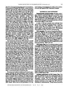
Introduction Polymerase chain reaction (PCR) method was a revolutionary innovation by Kary Mullis in the 1980s [1, 2]. Since this time, it has seen widespread use in biomedical research since it can detect and quantify small amounts of specific nucleic acid sequences. For example, small levels of messenger RNA (mRNA) can be quantified through the combination of reverse transcription (RT) to yield complementary DNA (cDNA) and PCR amplification to produce exponentially higher levels of these cDNA strands [3] (Fig. 1). In addition to increased levels of the amplified products (amplicons), the reliability and reproducibility of measurements between different laboratories are essential, especially if the method is to be performed in a clinical setting. This is critical for patient outcomes as well as for reducing healthcare costs since approximately one third of medical care budgets result from measurements and tests associated with diagnosis [4]. Quantitative PCR (qPCR) is a later development of the method that allows users to monitor the progress of a PCR reaction in real time [5]. In brief, the method uses a DNA-based sequence-specific
Paul C. Guest (ed.), Multiplex Biomarker Techniques: Methods and Applications, Methods in Molecular Biology, vol. 1546, DOI 10.1007/978-1-4939-6730-8_8, © Springer Science+Business Media LLC 2017
125
+
cDNA
Fig. 1 Schematic diagram of PCR
Nucleotides
3’
5’
+
Primer
5’
3’
3’
Denaturation
5’
5’
3’
Annealing
3’
5’
5’
3’
Elongation
3’
5’
5’
3’
Repeat
20-40 cycles
Repeat
126 Steve F.C. Hawkins and Paul C. Guest
Quantitative PCR
127
probe with a fluorescent reporter molecule at one end and a molecule that quenches this fluorescence at the other. The proximity of the reporter to the quench molecule prevents the detection of fluorescence and cleavage of the probe by the 5′ to 3′ exonuclease activity of Taq polymerase results in unquenched emission of fluorescence. Thus, the increase in the cDNA amplicon targeted by the reporter probe during each PCR cycle leads to a proportional increase in fluorescence due to cleavage of the probe and release of the reporter (Fig. 2). The available fluorescent reporter molecules include dyes that bind to double-stranded DNA such as SYBR® Green (Thermo Fisher Scientific; Waltham, MA, USA) or sequence specific probes like Molecular Beacons (Newark, NJ, USA), Scorpions (DxS Ltd), or TaqMan® Probes (Roche Molecular Diagnostics; Basel, Switzerland). As with standard PCR, qPCR is normally performed using a thermal cycler, which can rapidly heat and cool samples to allow the melting, annealing, and extension phases of replication. However in the case of qPCR, the thermocycler should also have the ability to illuminate each sample with specific wavelengths of light for the detection of the fluorescence emitted following excitation of the probe. PCR normally consists of a series of temperature changes that are repeated approximately 30 times. Each cycle consists of two or three steps. In the three step cycling approach, the first
Data Loading...