Myocardial Perfusion Scintigraphy in Acute Chest Pain: Current Status and Limitations
Patients with acute chest pain or symptoms suggestive of myocardial ischemia account for a large number of emergency department (ED) visits, approximately 7 million every year in the USA. Over 5 million of these patients are admitted to the hospital [1].
- PDF / 228,833 Bytes
- 6 Pages / 547.087 x 737.008 pts Page_size
- 57 Downloads / 332 Views
Myocardial Perfusion Scintigraphy in Acute Chest Pain: Current Status and Limitations
26
Priya Velappan and Assad Movahed
26.1
Contents
26.1 26.2 26.3 26.4 26.5 26.6 26.7 26.8
Introduction . . . . . . . . . . . . . . . . . . . . . . . . . . History . . . . . . . . . . . . . . . . . . . . . . . . . . . . . . Diagnostic Value of Acute Rest Myocardial Perfusion Imaging (ARMPI) . . . . . . . . . . . . Acute Rest Myocardial Perfusion Imaging and Outcome . . . . . . . . . . . . . . . . . . . . . . . . . Timing of Radionuclide Injection . . . . . . . . Cost Savings . . . . . . . . . . . . . . . . . . . . . . . . . . Conclusions . . . . . . . . . . . . . . . . . . . . . . . . . . Future Directions . . . . . . . . . . . . . . . . . . . . . References . . . . . . . . . . . . . . . . . . . . . . . . . . .
Introduction
299 300 300 301 301 301 302 302 303
Patients with acute chest pain or symptoms suggestive of myocardial ischemia account for a large number of emergency department (ED) visits, approximately 7 million every year in the USA. Over 5 million of these patients are admitted to the hospital [1]. However, only a minority of these patients have symptoms truly due to ischemic heart disease. The remainder does not require admission to the hospital and may cause a tremendous strain on limited economic resources. When the symptoms are classic and accompanied by diagnostic electrocardiographic changes, the diagnosis of acute coronary syndrome is straightforward. However, in patients with non-diagnostic ECG changes and classic or non-classic symptoms, appropriate decision making becomes more difficult. This dilemma has led to the exploration of newer diagnostic methods to appropriately risk-stratify patients in a cost-effective and time-sensitive manner. Myocardial perfusion imaging (MPI) provides a direct assessment of coronary blood flow and thus appears to be an optimal tool for identifying patients with acute coronary syndromes, who initially appear at low risk based on ECG or clinical characteristics. In acute ischemic syndromes, myocardial hypoperfusion occurs before the onset of left ventricular dysfunction, ECG changes, clinical symptoms and myocardial necrosis. Rest myocardial perfusion imaging becomes abnormal simultaneously with myocardial hypoperfusion. Radionuclide tracer uptake correlates in a linear fashion with blood flow in the range of resting myocardial perfusion and thus provides an accurate estimate of regional myocardial hypoperfusion. As MPI depends on flow abnormalities as well as myocardial necrosis, it can identify patients across the spectrum of acute coronary syndromes, including ischemia and infarction [1].
300
Priya Velappan and Assad Movahed
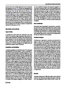
Fig. 26.1 Rest Tc-99m sestamibi images of a 52-year-old man with non-anginal chest pain and non-diagnostic electrocardiographic changes in the chest pain center. A relatively large inferoseptal defect is noted. (Reproduced with permission from [1])
26.2
History
Using rest MPI to identify patients with acute cor
Data Loading...