Nanoprobe Crosses Blood-Brain Barrier to Image and Treat Tumor
- PDF / 138,235 Bytes
- 2 Pages / 576 x 783 pts Page_size
- 86 Downloads / 266 Views
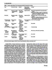
Nanoprobe Crosses Blood-Brain Barrier to Image and Treat Tumor Brain cancer is one of the hardest types of cancer to treat. Until now, no nanoparticle used for imaging has been able to cross the blood-brain barrier—an almost impenetrable barrier that protects the brain from infection—and specifically bind to braintumor cells. With current techniques, doctors inject dyes into the body and use drugs to temporarily open the blood-brain barrier, risking infection. Now, through use of a genetically engineered mouse model, M. Zhang and O. Veiseh of the University of Washington, J. Olson of Fred Hutchinson Cancer Research Center, R.G. Ellenbogen of the University of Wash ington and Seattle Children’s Hospital and Regional Medical Center, and their colleagues have been able to illuminate brain tumors by injecting fluorescent nanoparticles into the bloodstream that safely cross the blood-brain barrier. The nanoparticles remained in mouse tumors for up to five days and did not show any evidence of damaging the blood-brain barrier, according to results published in the August 1 issue of Cancer
Research (DOI: 10.1158/0008-5472.CAN09-1157; p. 6200). The nanoprobe is fabricated from an iron oxide nanoparticle coated with biocompatible polyethylene glycol–grafted chitosan copolymer. Crossing the bloodbrain barrier depends on the size of the particle, its lipid content, and the electric charge on the particle. The nanoparticle built by Zhang and her colleagues remains small in wet conditions. The particle was about 33 nm in diameter when wet, about a third the size of similar particles used in other parts of the body. To specifically target tumor cells, the research team used chlorotoxin, a small peptide isolated from scorpion venom that many research groups are exploring for its tumor-targeting abilities. On the nanoparticle’s surface, the researchers placed a near-infrared fluorophore for optical imaging, and binding sites that could be used for attaching other molecules. Results showed the nanoparticles improved the contrast in both magnetic resonance imaging and optical imaging, which is used during surgery. “Brain cancers are very invasive, differ-
CALL FOR PAPERS
ent from the other cancers. They will invade the surrounding tissue and there is no clear boundary between the tumor tissue and the normal brain tissue,” said Zhang, a professor of materials science and engineering. Being unable to distinguish a boundary complicates the surgery. Severe cognitive problems are a common side effect. “If we can inject these nanoparticles with infrared dye, they will increase the contrast between the tumor tissue and the normal tissue,” Zhang said. “So during the surgery, the surgeons can see the boundary more precisely. “We call it ‘brain tumor illumination or brain tumor painting,’” she said. “The tumor will light up.” Nano-imaging could also help with early cancer detection, Zhang said. Current clinical imaging techniques have a maximum resolution of 1 mm. Nanoparticles could improve the resolution by a factor of 10 or more, allowi
Data Loading...