Nondestructive magneto-optical imaging analysis of superconducting (Bi, Pb) 2 Sr 2 Ca 2 Cu 3 O x composites
- PDF / 217,481 Bytes
- 4 Pages / 612 x 792 pts (letter) Page_size
- 112 Downloads / 421 Views
MATERIALS RESEARCH
Welcome
Comments
Help
Nondestructive magneto-optical imaging analysis of superconducting (Bi, Pb)2 Sr2 Ca2 Cu3 Ox composites M. Turchinskaya, D. L. Kaiser, and A. J. Shapiro Materials Science and Engineering Laboratory, National Institute of Standards and Technology, Gaithersburg, Maryland 20899
G. N. Riley, Jr. and C. Christopherson American Superconductor Corporation, Two Technology Drive, Westborough, Massachusetts 01581 (Received 29 September 1995; accepted 22 February 1996)
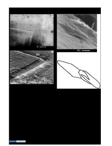
The superconducting filaments in (Bi, Pb)2 Sr2 Ca2 Cu3 OxyAg composite tapes have been imaged directly through the outer silver sheath of an unpolished tape by means of a magneto-optical imaging technique. The images reveal the morphology and alignment of the uppermost layer of filaments located as much as 112 mm below the unpolished tape surface, the depths of these filaments, and the homogeneity of the magnetic flux distribution within these filaments. These results demonstrate that the magneto-optical technique is a valuable nondestructive tool for analyzing (Bi, Pb)2 Sr2 Ca2 Cu3 Ox composite tapes. Silver-sheathed (Bi, Pb)2 Sr2 Ca2 Cu3 Ox (BSCCO) composites with transport critical current densities1 (Jct ) on the order of 104 Aycm2 are the leading high temperature superconducting materials for power applications. Recent magneto-optical (MO) imaging measurements taken under applied magnetic fields have provided considerable insight into the effect of microstructure on local current paths.2–6 To date, such measurements have all been conducted on polished sections of composite tape in order to obtain good physical contact between the BSCCO filaments and the optically active indictor film used in the MO experiments. With the same MO technique, we show here that the BSCCO filaments can be imaged directly through the outer silver sheath of an unpolished composite tape to disclose the positions of filaments located up to 112 mm below the tape surface and to reveal magnetic flux patterns developed within the composite under an applied magnetic field. These results are the first demonstration of the utility of the MO technique as a nondestructive means of evaluating BSCCO composites, pointing toward potential applications of MO imaging as a routine diagnostic tool for these materials. Two BSCCO multifilamentary composite tapes prepared by the powder-in-tube process1 were studied. One specimen with dimensions 0.29 mm 3 2.66 mm 3 7.0 mm (tape #1) contained 85 filaments embedded in a silver matrix, whereas the other specimen with dimensions 0.14 mm 3 2.45 mm 3 7.0 mm (tape #2) contained five filaments. The magnetic flux distributions in these two specimens were measured as a function of J. Mater. Res., Vol. 11, No. 7, Jul 1996
http://journals.cambridge.org
Downloaded: 14 Mar 2015
applied magnetic field (Ha ) at several temperatures by means of a MO technique employing a bismuth-doped yttrium iron garnet ferrimagnetic indicator film with inplane magnetic anisotropy.7 This MO technique has been used previously for investig
Data Loading...