Observation of Localized Corrosion of Ni-Based Alloys Using Coupled Orientation Imaging Microscopy and Atomic Force Micr
- PDF / 1,263,806 Bytes
- 6 Pages / 417.6 x 639 pts Page_size
- 38 Downloads / 262 Views
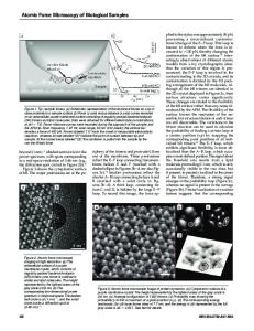
ABSTRACT We present a method for assessing the relative vulnerabilites of distinct classes of grain boundaries to localized corrosion. Orientation imaging microscopy provides a spatial map which identifies and classifies grain boundaries at a metal surface. Once the microstructure of a region of a sample surface has been characterized, a sample can be exposed to repeated cycles of exposure to a corrosive environment alternating with topographic measurement by an atomic force microscope in the same region in which the microstructure had been mapped. When this procedure is applied to Ni and Ni-based alloys, we observe enhanced attack at random grain boundaries relative to special boundaries and twins in a variety of environments. INTRODUCTION Advances in the engineering of grain boundaries in materials have been facilitated in recent years by the commercialization of a scanning electron microscope (SEM) technique, known as orientation imaging microscopy (OIMTM) [1,2,3], for automated indexing of electron diffraction backscattered Kikuchi patterns (EBSP). Using the nanometer-scale resolution of an Atomic Force Microscope (AFM), it is now possible to image the surface topography associated with the incipient stages of intergranular corrosion. Therefore, if the microstructure of a particular region is mapped with OIM, then subsequent alternation of corrosion and AFM imaging will reveal relative degree of attack associated with each grain boundary type during progressive stages of corrosion. In addition, it is possible to determine whether incipient stages of pitting corrosion are correlated in any way with microstructure. The work presented below is a demonstration of the coupled application of OIM and AFM to the corrosion of Ni-containing alloys in various corrosive environments.
81 Mat. Res. Soc. Symp. Proc. Vol. 586 ©2000 Materials Research Society
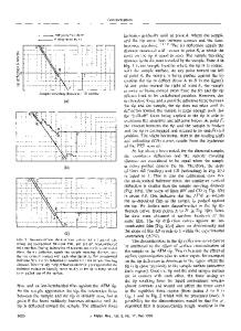
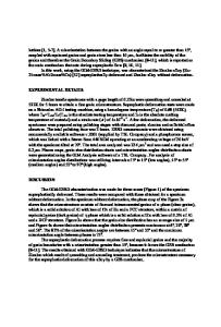
EXPERIMENT Metallographically-polished coupons of 316L stainless steel, Inconel 600, and commercially-pure, 201-grade Ni were first imaged with OIM to map their microstructures and grain-boundary orientations. [4] Indents were made in the samples to identify the locations mapped with OIM. The OIM set-up automatically acquires and processes EBSP's for determination of local orientations, misorientations, and microtexture. The interaction of the electron beam and the specimen generates an EBSP by the backscattering of electrons from favorably oriented crystal planes. Individual orientation measurements are made at discrete points on a sample; the locations of the points are defined by a grid of dimensions prescribed by the user (both in the width and height of the grid as well as the spacing between points on the grid). At each point in the grid, the backscattered Kikuchi diffraction pattern is captured, frame averaged and
automatically indexed. The three Euler angles that describe the orientation are recorded along with coordinates describing the position. Thus, images (or maps) can be generated by mapping the crystal orientation onto a color or gray scale and shading e
Data Loading...