Osteonecrosis Mimicking Bone Metastasis in Femoral Head on 18 F-FDG PET/CT: A Case Report
- PDF / 294,557 Bytes
- 4 Pages / 595.276 x 790.866 pts Page_size
- 92 Downloads / 329 Views
CASE REPORT
Osteonecrosis Mimicking Bone Metastasis in Femoral Head on 18F-FDG PET/CT: A Case Report Kyu-Ho Choi & Jin Kyoung Oh & Sung Hoon Kim & Ik Dong Yoo & Eun Kyoung Choi & Eun Ji Han
Received: 2 September 2010 / Accepted: 15 October 2010 / Published online: 11 November 2010 # Korean Society of Nuclear Medicine 2010
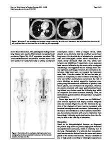
Abstract A 77-year-old woman underwent chemotherapy, radiotherapy, and brachytherapy for cervical cancer 9 years ago. On a follow-up 18F-fluorodeoxyglucose (FDG) PET/ CT image, focal FDG uptake was noted in a focal osteolytic lesion in the right femoral head. During magnetic resonance imaging, this lesion showed subchondral dark-signalintensity rim on T1-weighted image and double line sign on T2-weighted image, suggestive of osteonecrosis. The lesion was pathologically confirmed as osteonecrosis after surgery. This case demonstrates that osteonecrosis of the femoral head may demonstrate focal FDG uptake mimicking bone metastasis. Keywords Avascular necrosis . Femoral head . Bone metastasis . 18F-fluorodeoxyglucose . Positron emission tomography/computed tomography
Introduction 18
F- fluorodeoxyglucose (FDG) PET/CT scan demonstrates FDG accumulation in cancer cells with increased glucose metabolism and is widely used for staging, treatment response evaluation, residual tumor surveillance, and K.-H. Choi : J. K. Oh : S. H. Kim (*) : I. D. Yoo : E. K. Choi : E. J. Han Department of Radiology, The Catholic University of Korea, Seoul, Korea e-mail: [email protected] Present Address: S. H. Kim Department of Nuclear Medicine, Seoul St.Mary’s Hospital, The Catholic University of Korea, Seochogu Banpodong 505, 137-701 Seoul, Korea
detection of the recurrence of various malignancies. However, inflammatory diseases are known to show increased FDG uptake as well and can be mistaken for recurrence or metastasis. Focal FDG uptake in bone can readily be mistaken for bone metastasis [1–4]. Over the years, a few cases of osteonecrosis with focal uptake mistaken for bone metastasis or local recurrence have been reported [2, 3, 5]. On the other hand, there have been two studies on the FDG uptake of osteonecrosis [1, 6]. Osteonecrosis of the femoral head is a progressively debilitating lesion that is associated with a number of conditions including trauma, steroid use, alcohol abuse, and hypercholesterolemia; there are some clinical findings such as pain or limited motion [7, 8]. Imaging evaluation of osteonecrosis is usually done with radiography, magnetic resonance imaging (MRI), and bone scintigraphy. MRI continues to be the gold standard in diagnosing osteonecrosis of the femoral head [9]. We describe a case of osteonecrosis presenting as a focal FDG uptake combined with focal osteolytic lesion on corresponding CT images of FDG PET/CT, which mimicked bone metastasis in the femoral head.
Case Report A 77-year-old woman with cervical cancer who underwent chemotherapy, radiotherapy, and brachytherapy 9 years ago visited our hospital for surveillance and also complained of right hip pain for about 2 months. S
Data Loading...