Partial meniscectomy provides the favorable outcomes for symptomatic medial meniscus tear with an intact posterior root
- PDF / 705,409 Bytes
- 7 Pages / 595.276 x 790.866 pts Page_size
- 87 Downloads / 453 Views
KNEE
Partial meniscectomy provides the favorable outcomes for symptomatic medial meniscus tear with an intact posterior root Jae‑Young Kim1 · Seong‑Il Bin1 · Jong‑Min Kim1 · Bum‑Sik Lee1 · Sung‑Mok Oh1 · Won‑Joon Cho1 · Jae‑Hyung Lee1 Received: 17 December 2018 / Accepted: 16 July 2019 © European Society of Sports Traumatology, Knee Surgery, Arthroscopy (ESSKA) 2019
Abstract Purpose This study aimed to investigate the long-term outcomes of arthroscopic partial meniscectomy for medial meniscus tear (with intact posterior root) and to analyze the risk factors for treatment failure. Methods The records of 165 patients who underwent partial meniscectomy for medial meniscus tear with intact posterior root with a minimum 5-year follow-up were included. Modified Lysholm score and radiologic outcomes were compared between preoperative and latest follow-up periods. The cumulative Outerbridge grade of the medial compartment was defined as follows: 0–4, low chondral wear; 5–6, intermediate wear; or 7–8, high wear. Kaplan–Meier survival and Cox hazard regression analyses were performed to assess the survivorship after partial meniscectomy. Conversion to total knee replacement arthroplasty, high tibial osteotomy or a Lysholm score of 5°) at surgery and (b) concomitant additional procedures such as microfracture, synovectomy, or osteochondral transfer. The indications for APM were patients with confirmed medial meniscal tears who did not respond to at least 3 months of conservative treatment including non-steroidal, antiinflammatory drug medication, physical therapy, and muscle strengthening exercises and had persistent mechanical symptoms. A total of 165 patients who satisfied the above criteria were selected as candidates for this study. The average age of patients was 53.9 ± 9.2 years, and the average duration of symptoms was 25.5 ± 32.2 months. The average follow-up period was 103.6 ± 43.3 months (Table 1).
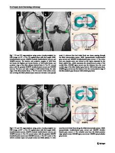
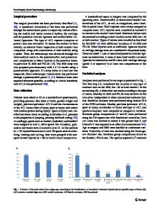
Assessment of tear morphology, cartilage status, arthritis, and clinical outcomes A single orthopedic knee surgeon performed arthroscopic surgery through anteromedial and anterolateral portals. Intraoperative findings, including meniscus lesions and cartilage status, were captured on digital imaging and transcribed into an electronic medical record. Meniscus tear
13
Knee Surgery, Sports Traumatology, Arthroscopy Table 1 Demographics of patients with medial meniscus tear with an intact posterior root Parameter
Value
Age (years) Male/female ratio Location Symptom duration (months) Follow-up period (months) BMI (kg/m2)
53.9 ± 9.2 43:122 Right: 89, Left: 76 25.5 ± 32.2 (5–120) 103.6 ± 43.3 (60–215) 25.2 ± 3.1
morphology was divided according to the ISAKOS classification as follows [2]: longitudinal-vertical, horizontal, radial, vertical flap, horizontal flap, and complex tear. The incidence of each tear type at meniscectomy and remnant rim width after meniscectomy was measured. Chondral wear at the time of APM was assessed using the Outerbridge classification (OB grade) [19]: grade 0 (normal cartilage), grade 1
Data Loading...