Transtibial fixation for medial meniscus posterior root tear reduces posterior extrusion and physiological translation o
- PDF / 1,331,977 Bytes
- 10 Pages / 595.276 x 790.866 pts Page_size
- 2 Downloads / 360 Views
KNEE
Transtibial fixation for medial meniscus posterior root tear reduces posterior extrusion and physiological translation of the medial meniscus in middle‑aged and elderly patients Yuya Kodama1,2 · Takayuki Furumatsu1 · Shin Masuda1 · Yoshiki Okazaki1 · Yusuke Kamatsuki1 · Yuki Okazaki1 · Takaaki Hiranaka1 · Shinichi Miyazawa1 · Masaharu Yasumitsu2 · Toshifumi Ozaki1 Received: 11 June 2019 / Accepted: 19 November 2019 © European Society of Sports Traumatology, Knee Surgery, Arthroscopy (ESSKA) 2019
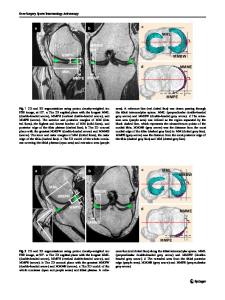
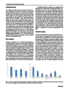
Abstract Purpose To investigate changes in meniscal extrusion during knee flexion before and after pullout fixation for medial meniscus posterior root tear (MMPRT) and determine whether these changes correlate with articular cartilage degeneration and short-term clinical outcomes. Methods Twenty-two patients (mean age 58.4 ± 8.2 years) diagnosed with type II MMPRT underwent open magnetic resonance imaging preoperatively, 3 months after transtibial fixation and at 12 months after surgery, when second-look arthroscopy was also performed. The medial meniscus medial extrusion (MMME) and the medial meniscus posterior extrusion (MMPE) were measured at knee 10° and 90° flexion at which medial meniscus (MM) posterior translation was also calculated. Articular cartilage degeneration was assessed using International Cartilage Research Society grade at primary surgery and second-look arthroscopy. Clinical evaluations included Knee Injury and Osteoarthritis Outcome Score, International Knee Documentation Committee subjective knee evaluation form, Lysholm score, Tegner activity level scale, and pain visual analogue scale. Results MMPE at 10° knee flexion was higher 12 months postoperatively than preoperatively (4.8 ± 1.5 vs. 3.5 ± 1.2, p = 0.01). MMPE at 90° knee flexion and MM posterior translation were smaller 12 months postoperatively than preoperatively (3.5 ± 1.1 vs. 4.6 ± 1.3, 7.2 ± 1.7 vs. 8.9 ± 2.0, p 5º, severe cartilage lesion [International Cartilage Research Society (ICRS) grade III or IV], and Kellgren–Lawrence (K–L) grade > II in radiographs. (b) Other than type II MMPRT. Among these 51 patients, 46 were diagnosed with type II MMPRT, under arthroscopic findings. Among the remaining five patients, one was diagnosed with type I MMPRT and four were diagnosed with type IV MMPRT. These five patients were excluded. Among the included 46 patients, 22 underwent open MRI preoperatively, as well as 3 and 12 months after surgery. Second-look arthroscopic evaluation was performed in all cases. This retrospective study analysed the changes in MMME and MMPE after transtibial fixation using open MRI and assessed cartilage degeneration using arthroscopic images and video recordings. We reviewed the patients’ medical records to determine age, sex, height, body weight, BMI, as well as preoperative and 3-month and 12-month postoperative clinical outcomes. The patient demographics are summarised in Table 1. Arthroscopic assessment of the cartilage lesions and anterior cruciate ligament (ACL) were performed using arthroscopic imag
Data Loading...