Pediatric rosette-forming glioneuronal tumor of the septum pellucidum
- PDF / 944,424 Bytes
- 4 Pages / 595.276 x 790.866 pts Page_size
- 58 Downloads / 290 Views
CASE REPORT
Pediatric rosette-forming glioneuronal tumor of the septum pellucidum Yazeed Al Krinawe 1 & Majid Esmaeilzadeh 1 & Christian Hartmann 2 & Joachim K. Krauss 1 & Elvis J. Hermann 1 Received: 10 February 2020 / Accepted: 6 March 2020 # Springer-Verlag GmbH Germany, part of Springer Nature 2020
Abstract Rosette-forming glioneuronal tumor (RGNT) is a rare tumor entity which has been reported mainly occurring in the fourth ventricle. It has been described as a benign lesion with limited extension into surrounding structures, including the cerebellar vermis, midbrain, and cerebral aqueduct. More recently, few cases involving also other midline structures have been documented as well. Here, we report about diagnosis and treatment of RGNT in the septum pellucidum in a pediatric patient which has not been described previously. A 7-year-old boy had a 3-week history of headache. Magnetic resonance imaging showed a solid mass in the septum pellucidum accompanied by hydrocephalus. The tumor was resected via a transcortical approach. Histological examination revealed the typical findings of a RGNT. At 2-year follow-up, there was no tumor recurrence, and clinical outcome was unremarkable. RGNT has to be considered in the differential diagnosis of pediatric midline tumors also outside of the fourth ventricle. Surgical resection is the first-line therapy which may result in beneficial outcome in the long term. The role of adjuvant therapy needs further definition since due to the rarity of this tumor entity, available data is very limited. Keywords Rosette-forming glioneuronal tumor . Septum pellucidum . Pediatric neurosurgery
Introduction Rosette-forming glioneuronal tumor (RGNT) is a rare tumor located primarily in the fourth ventricle, which was first described as a new tumor entity by Komori et al. in 2002 [1–3]. Young adults (mean age 33 years) have been mainly affected [4–6]. It usually presents as a benign lesion with limited extension into surrounding structures, including the cerebellar vermis, midbrain, and cerebral aqueduct. Rarely, cases manifesting in other locations in the central nervous system have been documented [7–10]. When surgical resection of the tumor is feasible, a generally favorable prognosis has been described. Yazeed Al Krinawe and Majid Esmaeilzadeh contributed equally to this work. * Yazeed Al Krinawe [email protected] 1
Department of Neurosurgery, Hannover Medical School, Carl-Neuberg Str. 1, 30625 Hannover, Germany
2
Department of Neuropathology, Institute of Pathology, Hannover Medical School, Hannover, Germany
Thus far, the occurrence of RGNT in the septum pellucidum has not been reported in a pediatric patient. Here, we describe our experience with the treatment and diagnosis of RGNT in the septum pellucidum in a 7-year-old boy.
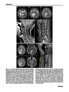
Case report A 7-year-old boy, who was born at term after an uncomplicated pregnancy and delivery, was referred with a 3-week history of headache. The past medical history revealed no intracranial infection, head injury, or previous surgery.
Data Loading...