Premalignant Lesions
Like other epithelial neoplasms, both hepatocellular carcinoma (HCC) and cholangiocarcinoma arising in the background of cirrhosis and chronic inflammatory biliary disorders are thought to originate from a premalignant stage.
- PDF / 1,770,236 Bytes
- 7 Pages / 595.28 x 790.87 pts Page_size
- 31 Downloads / 374 Views
Like other epithelial neoplasms, both hepatocellular carcinoma (HCC) and cholangiocarcinoma arising in the background of cirrhosis and chronic inflammatory biliary disorders are thought to originate from a premalignant stage. The histologic appearance of the precursor lesions of HCC has been the subject of extensive investigations and debate for many years. This probably results from the complex tridimensional structure of the hepatic plates and the architectural changes characteristic of cirrhosis, which result in a complex array of premalignant hepatocellular changes, clonal expansions, and nodular proliferations, with a blurred transition to overt malignancy. During the process of hepatocarcinogenesis,
12
several phenotypic changes arise, often designated as premalignant lesions, including hepatocellular cytologic changes, microscopic expansile foci (dysplastic foci, up to 1 mm), and macroscopic nodular lesions (dysplastic nodules). In contrast, biliary dysplasia is conceptually and morphologically similar to dysplasia of other “luminal” epithelium, and follows similar rules and criteria. Infiltration through the basal membrane marks the transition from in situ disease to invasive malignancy. Biliary premalignant lesions bear considerable similarities to their pancreatic counterparts, probably the result of their common embryologic origin.
A.W.H. Chan et al., Atlas of Liver Pathology, Atlas of Anatomic Pathology, DOI 10.1007/978-1-4614-9114-9_12, © Springer Science+Business Media New York 2014
171
172
12
Premalignant Lesions
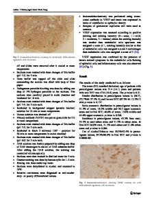
Fig. 12.1 Large cell change. These hepatocytes exhibit cellular and nuclear enlargement, nuclear pleomorphism and binucleation, prominent nucleoli, and a preserved nucleocytoplasmic ratio. Large cell change, formerly known as large cell dysplasia, now is believed to represent a pathophysiologic nuclear polyploidy, a degenerative senescent change occurring as a consequence of chronic liver injury, and in particular cholestasis. Large cell change is best regarded as a predictive marker associated with the risk of HCC rather than a genuine premalignant lesion. However, recent data suggest that large cell change, in some situations, may be a genuine premalignant lesion.
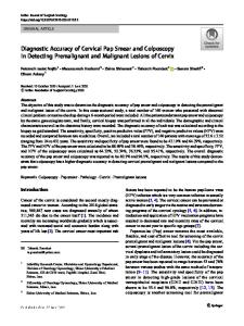
Fig. 12.3 Iron-free foci. Several hepatic nodules are free of haemosiderin deposition in a liver explant of hereditary haemochromatosis. Ironfree foci in hereditary haemochromatosis are shown to be more frequent in livers with HCC (50.0%) than in those without (8.3%) and to have a higher proliferative index and coexisting large cell change/small cell change (71.4%). HCCs in hereditary haemochromatosis not uncommonly are iron-free, and the sequential development from iron-free nodules to iron-free HCC in a recent rat model supports these foci as premalignant lesions. However, the underlying mechanisms of iron resistance in these foci and subsequent malignant transformation remain unknown.
Fig. 12.2 Small cell change. Hepatocytes exhibit decreased cell volume, minimal nuclear pleomorphism, increased nucleocytoplasmic rat
Data Loading...