Primary Esophageal CD30-Positive ALK-Positive Anaplastic Large Cell Lymphoma: A Case Report and Literature Review
- PDF / 318,707 Bytes
- 4 Pages / 595.276 x 790.866 pts Page_size
- 82 Downloads / 326 Views
BRIEF COMMUNICATION
Primary Esophageal CD30-Positive ALK-Positive Anaplastic Large Cell Lymphoma: A Case Report and Literature Review Ning Wu & Liewen Pang & Zhiming Chen & Yiqing Wang & Qinyun Ma & Gang Chen & Ji Chen & Jiechun Huang
Published online: 4 June 2010 # Springer Science+Business Media, LLC 2010
Abstract Purpose To introduce a case of primary esophageal CD30positive ALK-positive anaplastic large cell lymphoma (ALCL) and discuss its diagnosis and treatment. Methods Esophagectomy was done for a 37-year-old male with a submucosal lesion after a frozen section failed to give a definite diagnosis. Samples were sent for hematoxylin– eosin staining and immunohistochemical analysis. Six cycles of CHOP (cyclophosphamide, doxorubicin, vincristine, and prednisone) chemotherapy were given and the patient was followed up. Results The operation was uneventful. Postoperative pathologic and immunohistochemical examination yielded a diagnosis of primary CD30-positive and ALK-positive ALCL of the esophagus. The patient was in complete remission at the 14-month follow-up. Conclusions ALCL of the esophagus should be considered in the differential diagnosis of esophageal submucosal lesions. Biopsy through either esophagoscopy or surgical exploration, chemotherapy, and radiotherapy can be chosen for long-term survival. Keywords esophageal lymphoma . anaplastic large cell lymphoma . CD30-positive . ALK-positive
N. Wu : L. Pang (*) : Z. Chen : Y. Wang : Q. Ma : G. Chen : J. Chen : J. Huang Department of Cardiothoracic Surgery, Huashan Hospital, Fudan University, Shanghai 200040, People’s Republic of China e-mail: [email protected]
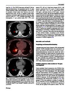
Clinical Summary In September 2008, a 37-year-old male was admitted to our department with a 3-month history of progressive dysphagia and a 10-kg weight loss. Physical examinations were normal. The results of blood routine and biochemistry tests were unremarkable. A submucosal mass was seen by esophagoscopy in the middle thoracic esophagus, 32 cm down from the incisors, but endoscopic biopsies failed to find any tumor cell. Endoscopic ultrasonography showed a submucosal neoplasm. Barium esophagogram revealed a 4×3×2 cm mid-esophageal mass with relatively smooth esophageal mucosa outline (Fig. 1a). The mass was visible on the computed tomography (CT) of the chest and was of soft-tissue density extending from just above the carina to just below the inferior pulmonary vein. Compression of the left and right bronchus was noted, whereas bronchoscopy excluded the possibility of bronchial mucosa involvement (Fig. 1b). A right thoracotomy was performed so as to obtain enough tissue for histologic analysis. The tumor involved approximately two thirds of the esophagus circumferentially, and was inseparable from the subcarinal nodes. After careful mobilization from both bronchi, a 2× 2×1 cm sample was sent for frozen section. The pathologic examination, however, was unable to indentify the exact type of malignancy, and therefore esophagectomy was performed with removal of adjacent enlarged lymph nodes. Cervi
Data Loading...