Primary merkel cell carcinoma clinically presenting as deep oedematous mass of the groin
- PDF / 2,434,987 Bytes
- 3 Pages / 595.232 x 841.846 pts (A4) Page_size
- 8 Downloads / 329 Views
274
June 28, 2010
Eur J Med Res (2010) 15: 274-276
© I. Holzapfel Publishers 2010
PRIMaRy MERkEl CEll CaRCInoMa ClInICally PREsEntIng as DEEP oEDEMatous Mass of tHE gRoIn t. gambichler 1, s. kobus 1, a. kreuter 1, u. Wieland 2, M. stücker 1 1 Department
of Dermatology, Ruhr-university Bochum, Bochum, germany,
2 Institute
of Virology, university of Cologne, Cologne, germany
Abstract Merkel cell carcinoma (MCC) is a relatively rare, polyomavirus associated, primary neuroendocrine carcinoma of the skin which is usually arising from dermal skin layers. However, the origin of MCC in the subcutaneous tissue is debatable. We report a 58-yearold female patient with an oedematous mass on her left groin that was firm in consistency and had no discoloration or other visible abnormality of the overlying skin. on histology and immunohistology the tumour was consistent with the diagnosis of MCC showing a predominant subcutanous growth pattern. Pelvic magnetic resonance tomography revealed a tumour conglomerate reaching from the subcutis of the left groin to the left paraaortal and parailiacal region indicating widespread lymphogenic metastisation. Despite complete medical work-up no other MCC primary could be detected. In conclusion, predominant subcutaneous growth pattern as well as tumour localization in the groin are uncommon features of MCC. MCC showing the aforementioned features may be associated with significant delay of diagnosis and therefore represents an unfavourable prognostic factor.
IntRoDuCtIon Merkel cell carcinoma (MCC) is a relatively rare, polyomavirus associated, primary neuroendocrine carcinoma of the skin usually occurring on sun-exposed areas in elderly patients [1]. the typical clinical manifestation of MCC is that of a firm, nontender, cutaneous nodule with red-, purple-, or violet-coloured skin. It may display aggressive biological behaviour and can grow rapidly, metastasising to the regional lymph nodes and other organs such as liver, bones, lungs and brain [1, 2]. In this report, we describe an unusual case of MCC clinically presenting as oedematous mass of the left groin.
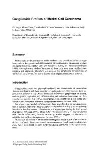
CasE REPoRt a 59-year-old woman presented with a painless mass of about 3 months duration in the left inguinal and vulval area. she had a 4-week history for a common iliac vein thrombosis of the left leg. the oedematous mass of the left groin was firm in consistency and had no discoloration or other visible abnormality of the overlying skin, except for a slight ‘peau d’orange’ ap-
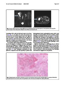
pearance (fig. 1a). Pelvic magnetic resonance tomography revealed a tumour conglomerate reaching from the subcutis of the left groin to the left paraaortal and parailiacal region indicating widespread lymphogenic metastisation (fig. 1b). neoplasms of the uro-genital regions were excluded by urologists and gynaecologists, respectively. a deep incisional skin biopsy was performed. Histological examination revealed a diffuse growth pattern with polygonal-epitheloid tumour cells of “naked” nuclei, coarse chromatin, and frequent mitotic features (fig
Data Loading...