Pure laparoscopic liver resection for giant liver hemangioma with extrahepatic growth based on preoperative 3-dimensiona
- PDF / 930,872 Bytes
- 4 Pages / 595.276 x 790.866 pts Page_size
- 77 Downloads / 370 Views
CASE REPORT
Open Access
Pure laparoscopic liver resection for giant liver hemangioma with extrahepatic growth based on preoperative 3-dimensional simulation: A case report Yuichiro Okumura1, Takehiro Noda1, Hidetoshi Eguchi1* , Takehiko Hanaki1, Yoshifumi Iwagami1, Hirofumi Akita1, Tadafumi Asaoka1, Kunihito Gotoh1, Shogo Kobayashi1, Koji Umeshita2, Masaki Mori1,3 and Yuichiro Doki1
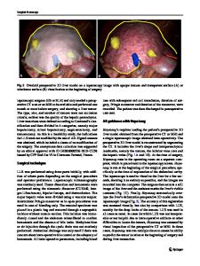
Abstract Background: Performing laparoscopic liver resection for giant hemangiomas is challenging, and careful preoperative planning is essential. Controlling intraoperative bleeding and handling surgical instruments within a limited workspace is necessary. Case presentation: In the present case, the patient was a 38-year-old woman diagnosed with a 16-cm giant liver hemangioma in segment 5/6, with extrahepatic growth. Preoperative three-dimensional simulations for port placement and the laparoscopic view from the left upper abdomen were performed to complete the pure laparoscopic liver resection. The laparoscopic resection was then safely performed on the same way. Conclusions: Pure laparoscopic resection could be applied to giant hemangiomas with extrahepatic growth, and the preoperative three-dimensional simulation of port placement and the laparoscopic view might be helpful when the intraabdominal workplace is restricted. Keywords: Giant hemangioma, Laparoscopic liver resection, 3D simulation
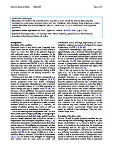
Introduction Cavernous hemangiomas are the most common type of benign liver tumors, and the majority of cavernous hemangiomas are incidentally detected by imaging studies performed for other reasons [1]. A recent national survey reported that liver hemangiomas measuring > 10 cm would be candidates for surgery in patients with symptoms [2]. Compared with open liver resection, laparoscopic liver resection (LLR) has many advantages including shorter hospital stay, less blood loss, and earlier postoperative recovery [3]. The current indication for LLRs is a solitary tumor with a ≤ 5-cm diameter [4]. LLR for giant hemangiomas > 10 cm is challenging due to insufficient space for manipulation and bleeding risk [2, 3]. Here, we present a case with a symptomatic giant hemangioma 16 cm in * Correspondence: [email protected] 1 Department of Gastroenterological Surgery, Graduate School of Medicine, Osaka University, 2-2, Yamadaoka E-2, Suita, Osaka 565-0871, Japan Full list of author information is available at the end of the article
diameter that was successfully resected by LLR after utilizing three-dimensional (3D) simulation technology to preoperatively plan port placement and the laparoscopic view.
Case presentation A young woman was referred to our hospital for a hepatic mass, which presented with epigastric pain. The patient had no notable past medical history. She did not smoke or abuse alcohol. Her physical examination revealed slight epigastric tenderness. Contrast-enhanced computed tomography (CT) demonstrated a 9-cm mass in segments 5/6 of the liver, with extrahepatic growth. The radiological findings were compatible with ca
Data Loading...