A middle-aged man presenting with progressive ataxia and pendular nystagmus: a delayed onset post-stroke movement disord
- PDF / 769,924 Bytes
- 3 Pages / 595.276 x 790.866 pts Page_size
- 63 Downloads / 222 Views
LETTER TO THE EDITOR
A middle‑aged man presenting with progressive ataxia and pendular nystagmus: a delayed onset post‑stroke movement disorder Byoung June Ahn1 · Mina Lee2 · Hyunjin Ju2 · Kayeong Im2 · Kyum‑Yil Kwon2 Received: 13 January 2020 / Accepted: 6 April 2020 © Belgian Neurological Society 2020
Introduction Hypertrophic olivary degeneration (HOD) is a rare neurological condition, which is defined as a secondary degenerative process associated with focal lesions to the Guillain–Mollaret triangle of the dentato-rubro-olivary pathway. The etiologies of HOD include hemorrhage, ischemia, tumors, and demyelination [1, 2]. Classically, patients with HOD show characteristic palatal tremors combined with a variable degree of other neurological signs. Herein, we describe a middle-aged man with HOD that developed 6 months after dorsal pontine hemorrhage, who presented with cerebellar ataxia and pendular nystagmus but not palatal tremor.
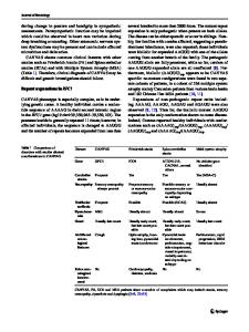
Case report A 55-year-old man was admitted to our hospital for evaluation of progressive gait difficulty. Fourteen months previously, the patient suddenly developed severe postural instability due to hemorrhagic stroke in the pontine tegmentum (Fig. 1a). He had no history of any medical illness and hypertension was diagnosed at that time. Through comprehensive rehabilitation therapy for a half year, the patient nearly recovered from his postural instability and could walk independently. However, over the following 8 months, his Electronic supplementary material The online version of this article (https://doi.org/10.1007/s13760-020-01354-x) contains supplementary material, which is available to authorized users. * Kyum‑Yil Kwon [email protected] 1
Department of Neurology, Soonchunhyang University Gumi Hospital, Soonchunhyang University College of Medicine, Gumi, Korea
Department of Neurology, Soonchunhyang University Seoul Hospital, Soonchunhyang University College of Medicine, 59 Daesagwan‑ro, Yongsan‑gu, Seoul 04401, Republic of Korea
2
gait disturbance worsened slowly and he became wheelchair bound. The neurological examination revealed a moderate of limb ataxia, which was more prominent in the left extremities than in the right extremities. Sinusoidal oscillation in his eyes was observed but palatal tremor was not noted. Cognitive function tests, as well as motor and sensory examinations, were unremarkable. The patient’s routine laboratory tests were normal. T2-weighted magnetic resonance imaging (MRI) revealed high signal intensity in the bilateral inferior olivary nucleus (ION), which was more predominant in the right side than in the left side (Fig. 1b). Video electronystagmography showed pendular nystagmus of 2.5 Hz frequency, which was mainly horizontally directed and more severe in the left eye than the right eye (Fig. 1c). Further evaluations, including a cerebrospinal fluid study and detailed tests for paraneoplastic syndrome, yielded unremarkable findings. The patient became bedridden after several months and we have followed him for one and a half year
Data Loading...