A new approach to characterization of the transition temperatures of thin film NiTi shape memory alloys
- PDF / 307,953 Bytes
- 6 Pages / 612 x 792 pts (letter) Page_size
- 35 Downloads / 326 Views
Jianxin Zhang and Clive J. Roberts Laboratory of Biophysics and Surface Analysis, School of Pharmaceutical Science, University of Nottingham, Nottingham NG7 2RD, United Kingdom (Received 25 April 2003; accepted 8 March 2004)
A novel approach in determining the transition temperatures of NiTi shape memory alloys was investigated and compared with conventional techniques. The technique is based on microthemal analysis using a scanning thermal microscope (SThM). In particular, this method has the potential to allow the transformation temperatures of thin films to be investigated in situ. Thin film shape memory alloys have potential applications, such as microactuators, where conventional analysis techniques are either not directly applicable to such samples or are difficult to perform.
I. INTRODUCTION
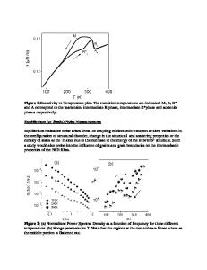
Near equiatomic nickel titanium (NiTi) shape memory alloys have been used in a wide range of applications since their rediscovery in 19621 due to their ability to recover from up to 10% strain, with recovery stresses approaching 400 MPa. One common application is as actuators, in which NiTi elements produce not only a greater force than competing bimetals, but over a larger range and with a narrower window of temperature actuation; consequently, they have been used for many applications such as window openers, mixing taps, and airconditioning flow controllers.2,3 More recently, interest in NiTi thin films as potential microactuators, that is, for micro-valves and micro-positioners4–6 has generated interest. Crucial to all these devices is the control and accurate determination of the martensitic to austenitic transformation temperature. The martensitic to austenite transformation temperatures of bulk NiTi can be readily assessed by bulk methods such as differential scanning calorimetry (DSC) and themomechanical methods, neither of which are readily adaptable to thin coatings due to a requirement of sufficient sample mass or limitations imposed by the substrate. Sheet resistance measurements or thermal x-ray diffraction (XRD) may be used on relatively large areas of thin film coatings, but this does not meet the smallscale requirements of the electronics industry. Scanning thermal microscopy (SThM) offers a new method that
a)
Address all correspondence to this author. e-mail: [email protected] DOI: 10.1557/JMR.2004.0240 1762
J. Mater. Res., Vol. 19, No. 6, Jun 2004
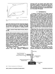
has the potential to offer in situ determination of these transformation temperatures at the micrometer level. The basic concept of the SThM is to replace the tip of a conventional atomic force microscope with a thermal sensor, allowing it to continue to perform in its conventional role by providing topographical information from a sample under investigation, while simultaneously acting as a small heater and resistance thermometer to obtain local thermal analysis with micrometer spatial resolution. In 1994, Pylkki et al.7 pioneered the use of an etched Wollaston wire thermal tip that combined the features of both heater and thermocouple into a
Data Loading...