Automated diffraction tomography combined with electron precession: a new tool for ab initio nanostructure analysis
- PDF / 2,983,063 Bytes
- 13 Pages / 612 x 792 pts (letter) Page_size
- 33 Downloads / 293 Views
1184-GG01-05
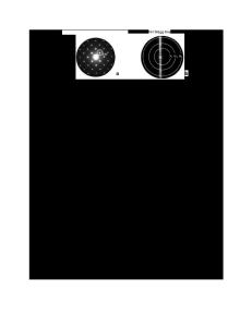
Automated diffraction tomography combined with electron precession: a new tool for ab initio nanostructure analysis Ute Kolb*, Tatiana Gorelik, Enrico Mugnaioli Institut für Physikalische Chemie, Johannes Gutenberg-Universität Mainz, Welderweg 11, D55099 Mainz, Germany ABSTRACT Three-dimensional electron diffraction data was collected with our recently developed module for automated diffraction tomography and used to solve inorganic as well as organic crystal structures ab initio. The diffraction data, which covers nearly the full relevant reciprocal space, was collected in the standard nano electron diffraction mode as well as in combination with the precession technique and was subsequently processed with a newly developed automated diffraction analysis and processing software package. Non-precessed data turned out to be sufficient for ab initio structure solution by direct methods for simple crystal structures only, while precessed data allowed structure solution and refinement in all of the studied cases.
INTRODUCTION The rapidly developing nanotechnology urgently needs analytical tools to characterize nano-volumes. The crystalline structure of a material is a principal key for understanding its properties and therefore is the most desired piece of information. Well developed methods for structure analysis by X-ray single crystal diffraction are established and routinely used in many laboratories. Single crystal X-ray analysis requires crystals with a size of at least about 1 mm3. Powder X-ray diffraction can access significantly smaller crystals but indexing and subsequent structure solution is often problematic due to peak overlap, the presence of additional phases, and a preferred orientation. Furthermore, the problem of peak overlap is particularly enhanced due to crystal-size driven peak broadening for nanocrystalline materials. High resolution transmission electron microscopy (HRTEM) is traditionally used for nanostructural investigations. Extrapolating three-dimensional (3D) structural information from images requires special tomographic techniques, which are typically not optimized for electron beam sensitive materials, such as inorganic complex structures (i.e. zeolites) and organic crystals, which cannot sustain the high electron dose needed to collect the data from nano-volumes. While strong efforts were dedicated in recent years for the construction of aberration correctors in order to achieve sub-Ångstrom resolution in imaging [1], the usage of the equivalent information in reciprocal space, already providing such a resolution and easily available in any transmission electron microscope (TEM), is not so well developed. Electron diffraction can probe volumes down to 20-30 nm in diameter, delivering 3D sub-Ångstrom structural information with good signal-to-noise ratio. Nevertheless, there are only a few software packages using low index zone axis patterns [2] and no hardware or formalism for dealing with patterns that were recorded though a tilt around an arbitrary axis available so fa
Data Loading...