Balint-Holmes syndrome due to stroke following SARS-CoV-2 infection: a single-case report
- PDF / 225,782 Bytes
- 3 Pages / 595.276 x 790.866 pts Page_size
- 31 Downloads / 264 Views
COVID-19
Balint-Holmes syndrome due to stroke following SARS-CoV-2 infection: a single-case report Francesco Panico 1
&
Angela Arini 2 & Pierluigi Cantone 2 & Claudio Crisci 2 & Luigi Trojano 1
Received: 17 September 2020 / Accepted: 24 October 2020 # Fondazione Società Italiana di Neurologia 2020
Dear Editor-in-Chief, Balint-Holmes’ syndrome (BHS) is a neuropsychological condition related to bilateral lesions in the parieto-occipital cortex, characterized by simultanagnosia, ocular apraxia, and optic ataxia [1, 2]. Simultanagnosia is the inability to detect multiple visually presented objects (dorsal type) and to recognize the individual parts of a multipart object (ventral type). Ocular apraxia is an impairment in saccade initiation and visual pursuit, despite unrestricted ocular movements. Optic ataxia is the difficulty in performing movements directed to visual objects, in the absence of primary sensory and motor disorders. BHS has been described mainly after cerebrovascular events, but also in relation to HIV encephalopathy, carbon monoxide poisoning, or posterior cortical atrophy [1, 2]. Here we report a patient showing BHS following bilateral parieto-occipital damage due to a stroke after SARS-CoV-2 infection. A 55-year-old male teacher, with systemic arterial hypertension under pharmacological treatment (ramipril and amlodipine) and mild hypercholesterolemia, suddenly developed fever, cough, dyspnea, ageusia, and anosmia in March 2020. After detection of SARS-CoV-2 viral nucleic acid by a nasopharyngeal swab, the diagnosis of coronavirus disease 2019 (COVID-19) was made. In a few days, the patient was admitted to intensive care unit for worsening of respiratory distress.
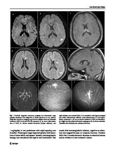
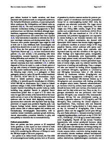
At discharge from intensive care unit, one month later (April 16, 2020), DG presented with tetraplegia and vision loss. A magnetic resonance scan showed temporo-parietooccipital cortico-subcortical lesions in the right hemisphere and parieto-occipital cortico-subcortical lesions in the left hemisphere (Fig. 1) which were compatible with a bilateral stroke in the territory of the inferior division of the middle cerebral artery. When the patient was transferred to a rehabilitation unit (May 2020), he was alert and cooperative, and showed mild bilateral hyposthenia with hyporeflexia, mild tactile sensory loss, and left homonymous hemianopia. The neuropsychological examination at bedside revealed left spatial neglect (Fig. 2a) associated with word finding difficulties and mild executive dysfunction.
First authorship is shared by Francesco Panico and Angela Arini * Francesco Panico [email protected] 1
Department of Psychology, University of Campania “Luigi Vanvitelli”, Viale Ellittico 31, 81100 Caserta, Italy
2
Clinic Center Rehabilitation Institute, Viale Maria Bakunin 171, 80126 Naples, Italy
Fig. 1 Axial brain T1-MR scan showing the wide right temporo-parietal and the left parietal lesions
Neurol Sci
Fig. 2 a Patient’s performance in a line cancellation test. b Percentage of correct responses in a task to assess op
Data Loading...