Cardiac Muscle
Cardiac muscle belongs to the striated types of muscle containing the same arrangements of myofilaments as skeletal muscle. However, unlike in skeletal muscle, no syncytia are built but cylindrical muscle cells containing one large cube-shaped nucleus in
- PDF / 1,390,565 Bytes
- 2 Pages / 595.28 x 790.87 pts Page_size
- 81 Downloads / 330 Views
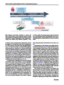
anes are visible. Both cells are covered by a basal lamina and show multiple caveolae-like vesicles close to the cell surfaces. Triads are not as regular and prominent as in skeletal muscle, and diads are often formed instead of triads. However, anastomozing networks of the sarcoplasmic reticulum surround the myofibrils, to be seen in both panels A and B (arrowheads). Mitochondria (M) are numerous; they show densely packed cristae and occupy the cytoplasm between the myofibrils where also glycogen is stored. The energy storing, energy releasing, and energy recapturing structures and organelles, glycogen particles and mitochondria, are clearly visible adjacent to the myofibrils where the energy is used for contraction. Three-dimensional studies of the membrane systems for Ca2+ signaling in cells of the mouse myocardium revealed detailed insights into the structures of diads, and showed frequent physical links between the outer mitochondrial membrane and the sarcoplasmic reticulum and T-tubules. The area indicated by a rectangle in panel B is shown at higher magnification in the inset. The section leads through an A-band, where thick myosin and thin actin filaments overlap. Each thick myosin filament is located within the center of a hexagonal array of thin actin filaments. Such a unit is encircled. For adaptation according to the physiological demands, sensing of biomechanical signals is required involving the interfaces between myofibrils and the plasma membrane at the cell-to-cell junctions within the intercalated disks and the cell-matrix junctions at the costameres (cf. Fig. 169). It is assumed that the giant protein titin has a main role as a biomechanical sensor.
References Franzini-Armstrong C, Protasi F, Ramesh V (1998) Comparative ultrastructure of Ca2+ release units in skeletal and cardiac muscle. Ann N Y Acad Sci 853:20 Hayashi T, Martone ME, Yu Z, Thor A, Doi M, Hoist MJ, Ellisman MH, Hoshijima M (2008) Three-dimensional electron microscopy reveals new details of membrane systems for Ca2+ signaling in the heart. J Cell Sci 122:1005 Miller MK, Granzier H, Ehler E, Gregorio CC (2004) The sensitive giant: the role of titin-based stretch sensing complexes in the heart. Trends Cell Biol 14:119 Severs NJ (2000) The cardiac muscle cell. Bioessays 22:188
Magnification:×35,500 (A), ×114,000 (B), ×273,600 (inset) M. Pavelka, J. Roth, Functional Ultrastructure: Atlas of Tissue Biology and Pathology, Third Edition, DOI 10.1007/978-3-7091-1830-6_27, © Springer-Verlag Vienna 2015
350
Figure 174
351
Data Loading...