Do different cone beam computed tomography exposure protocols influence subjective image quality prior to and after root
- PDF / 506,768 Bytes
- 9 Pages / 595.276 x 790.866 pts Page_size
- 93 Downloads / 215 Views
ORIGINAL ARTICLE
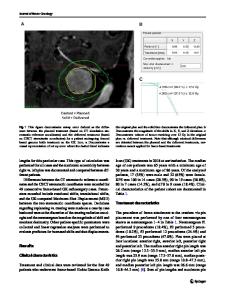
Do different cone beam computed tomography exposure protocols influence subjective image quality prior to and after root canal treatment? Andy Wai Kan Yeung 1 & Basak Harper 1 & Chengfei Zhang 2 & Prasanna Neelakantan 2 & Michael M. Bornstein 1,3 Received: 19 November 2019 / Accepted: 11 August 2020 # The Author(s) 2020
Abstract Objectives The current study aimed to evaluate different CBCT exposure protocols and influencing factors affecting the subjective image quality of scans taken for endodontic indications. Materials and methods Twelve extracted teeth, comprising of two sets of maxillary molars, premolars, canines and incisors, mandibular premolars, and molars, were endodontically treated, and either received a fiber or metal post. The teeth were scanned by CBCT imaging before and after root canal treatment, and after post insertion. Each scan was performed thrice, using an ultra low dose (ULD), standard (SM), and high-resolution mode (HR), respectively. Twelve observers—4 endodontists, 4 periodontists, and 4 radiologists— assessed the subjective image quality using visual analogue scales (VAS). Potential influencing factors were evaluated including acquisition mode, observer specialty, stage of treatment, type of post, and type of tooth, using one-way ANOVA and T test. Results Teeth scanned with the ULD had the highest average VAS score (72.5), followed by HR (70.2), and SM (69.0) for values pooled from all teeth and observers. CBCT acquisition mode was not a significant influencing factor on the VAS scores. Observer specialty, stage of treatment, type of post, and type of tooth were significant influencing factors. Conclusions Based on the present in vitro data, a low-dose CBCT mode seems not to negatively affect the perception of image quality. Clinical relevance The findings from this in vitro study demonstrate that a low-dose CBCT mode might have potential for diagnostics prior to or following endodontic treatment. Keywords Cone beam computed tomography . Dose optimization . Root canal treatment . Image quality . Low dose
Introduction The pitfalls of two-dimensional (2D) radiographic imaging in endodontics are well known [1]. For diagnostic purposes, conventional radiographs may have limitations, such as to identify all root canals including blocked canals, evaluate the morphology of tooth and root canal systems, localize broken instruments, and assess the relationship between the roots and
* Michael M. Bornstein [email protected] 1
Oral and Maxillofacial Radiology, Applied Oral Sciences and Community Dental Care, Faculty of Dentistry, The University of Hong Kong, Hong Kong SAR, China
2
Endodontology, Faculty of Dentistry, The University of Hong Kong, Hong Kong SAR, China
3
Department of Oral Health & Medicine, University Center for Dental Medicine Basel UZB, University of Basel, Basel, Switzerland
the maxillary sinus or the mandibular canal [2]. Detection of cracks/fractures is also challenging using conventional radiographs, when the fracture lines are along the dire
Data Loading...