Early laser photocoagulation for extensive retinal avascularity in infants with incontinentia pigmenti
- PDF / 1,493,673 Bytes
- 8 Pages / 595.276 x 790.866 pts Page_size
- 58 Downloads / 285 Views
CLINICAL INVESTIGATION
Early laser photocoagulation for extensive retinal avascularity in infants with incontinentia pigmenti Shiro Nakao1 · Sachiko Nishina1 · Shin Tanaka1 · Tomoyo Yoshida1 · Tadashi Yokoi1 · Noriyuki Azuma1 Received: 7 December 2019 / Accepted: 16 July 2020 © Japanese Ophthalmological Society 2020
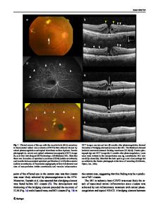
Abstract Purpose To describe the clinical features and treatment outcomes of severe retinopathy in eyes with incontinentia pigmenti (IP) of infants within a few months of birth. Study design Retrospective clinical study. Methods Six eyes of three patients (6-day-old girl, 5-month-old girl, and 14-day-old boy) with IP were examined and treated under general anesthesia. Ophthalmologic examinations were performed including images from wide-angle fluorescein angiography (FA), swept-source optical coherence tomography (OCT), and OCT angiography (OCTA). Results Ophthalmoscopy showed prominent vascular tortuosity in five eyes, retinal hemorrhages in four eyes, and incomplete vascular development in two eyes. FA showed extensive avascularity including the posterior pole of the retina in all cases except one eye. Prompt and intensive laser photocoagulation stabilized the pre-proliferative severe retinopathy in five eyes; however, foveal structure and vessel anomalies were detected in three of six eyes by OCT and two of five eyes by OCTA. Conclusion Severe retinopathy in the neonatal period and infancy was present not only in the periphery but also in the posterior pole including the fovea, which might be related to retinal vascular maldevelopment. It is, therefore, recommended that wide-angle fundus FA examination be performed in the early postnatal period to detect early signs of severe retinopathy in infants with IP. Keywords Incontinentia pigmenti · Retinopathy · Laser photocoagulation · Fluorescein angiography · Optical coherence tomography
Introduction Incontinentia pigmenti (IP), known as Bloch-Sulzberger syndrome, was described first in 1906 by Garrod [1]. A rare X-linked dominantly inherited syndrome mostly seen in females yet fatal in males, has an incidence of 1 in 40,000 newborns; in most cases, IP results from mutations in the IKBKG gene, responsible for impaired signaling of nuclear Corresponding Author: Noriyuki Azuma Electronic supplementary material The online version of this article (https://doi.org/10.1007/s10384-020-00768-7) contains supplementary material, which is available to authorized users. * Noriyuki Azuma azuma‑[email protected] 1
Laboratory for Visual Science, Department of Ophthalmology, National Center for Child Health and Development, 2‑10‑1 Okura Setagaya‑ku, Tokyo, Japan
factor kappa B, a critical transcription factor that up-regulates the immune system and prevents apoptosis [2]. Female infants with IP develop characteristic abnormalities of the skin, central nervous system (CNS), bones, hair, teeth, and eyes. Approximately 25–77% of cases are associated with ophthalmic manifestations, which primarily occur in the retina, such as vascular occlusion, neovascularizat
Data Loading...