Elastic Modulus and Mineral Density of Dentine and Enamel in Natural Caries Lesions
- PDF / 277,854 Bytes
- 6 Pages / 612 x 792 pts (letter) Page_size
- 101 Downloads / 315 Views
L5.15.1
Elastic Modulus and Mineral Density of Dentine and Enamel in Natural Caries Lesions Amanpreet K. Bembey1, Michelle L. Oyen2, Ching-Chang Ko2, Andrew J. Bushby1 and Alan Boyde3. 1
Department of Materials, Queen Mary, University of London, London E1 4NS, UK University of Minnesota, Minneapolis, MN 55455 3 Dental Institute, Queen Mary, University of London, London E1 1BB, UK 2
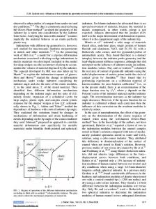
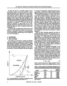
ABSTRACT Dental tissues have been reported to show a considerable decrease in both their mineral content and mechanical properties in carious lesions. The changed properties of dentine and enamel have been shown to be dependent on crystal size and not only mineral content [1], although the connectivity between the mineral crystals has been overlooked. Teeth with carious lesions were sectioned, embedded in polymethylmethacrylate (PMMA) and diamond polished. Nanoindentation and quantitative backscattered electron imaging were used to determine relationships between the elastic modulus and mineral density of sound and carious regions within dentine and enamel. The changes in elastic modulus with decreased mineralization for dentine and enamel could not be explained by simple composite mechanics expressions relating elastic modulus and mineral volume fraction. Finite element modeling of dentine and enamel as a two-phase composite material at the ultrastructure level were used to demonstrate how changes in the mineral phase connectivity can produce changes in the elastic modulus. Tissue models for enamel, in which the mineral phase is both the major component of the structure (~ 85% by volume) and highly interconnected, were consistent with the modulus of sound enamel. The drastic change in enamel modulus with a relatively small change in mineral volume fraction could be modeled as a decrease in mineral phase connectivity at nearly constant volume fraction. The more gradual trend in the dentine data was also consistent with a structure that is initially highly connected in the mineral phase, consistent with the known structure of dentine, and for which the change in modulus is more directly related to changes in mineral content than mineral connectivity. INTRODUCTION Mineralized tissues are composite materials with a mineral carbonated hydroxyapatite (HA) phase and an organic hydrated phase, largely collagen in the calcified connective tissues like bone and dentine, but there is no collagen in enamel. For composite materials with large modulus mismatch between the component phases, a wide range of modulus values can be achieved at constant composition (i.e. mineral volume fraction) by altering the geometrical structure and arrangement of the component phases. The precise arrangement of the component phases at the ultrastructural level is unclear for mineralized tissues such as teeth, and there is associated uncertainty with the changes in tissue composition and structure due to disease processes such as dental caries. Although there is certainly a loss of mineralization in caries, the way that this affects the structure of the component pha
Data Loading...