Heavy and light chain (AHL)-type cardiac amyloidosis: first histopathologic-proven case illustrating involvement of the
- PDF / 7,757,589 Bytes
- 6 Pages / 595.276 x 790.866 pts Page_size
- 88 Downloads / 284 Views
BRIEF REPORT
Heavy and light chain (AHL)-type cardiac amyloidosis: first histopathologic-proven case illustrating involvement of the heart Miroslav Sekulic 1
&
Simona Pichler Sekulic 1 & Astrid Weins 2
Received: 9 March 2020 / Revised: 23 April 2020 / Accepted: 28 April 2020 # Springer-Verlag GmbH Germany, part of Springer Nature 2020
Abstract Cardiac amyloidosis is most commonly comprised of either a monoclonal immunoglobulin or transthyretin; however, in practice, detailing of the former beyond light chain restriction is not typically performed. We present briefly the case of an 80-year-old man with concern for cardiac amyloidosis and a subsequent endomyocardial biopsy revealing significant deposition of amorphous Congo red-positive material. By immunofluorescence microscopy, the amyloidogenic material showed positive expression for IgG heavy chain and kappa light chain, with negative staining for IgM and IgA heavy chains and lambda light chain supporting a diagnosis of heavy and light chain (AHL)-type amyloidosis. Immunofluorescence staining for the IgG heavy chain subclasses supported and further classified the patient’s AHL-type cardiac amyloidosis as being IgG4/kappa restricted. The presented case is the first to illustrate AHL-type cardiac amyloidosis via sampling of heart tissue. Keywords Cardiac amyloidosis . AHL-type amyloidosis . Immunofluorescence microscopy . IgG heavy chain subclasses
Introduction
Case report
The most common types of amyloidosis to involve the heart are those derived from monoclonal immunoglobulin and transthyretin [1–3]. Although the most commonly reported type of cardiac amyloid when derived from a monoclonal immunoglobulin is comprised of a restricted immunoglobulin light chain (AL-type), amyloid in general can also be composed of immunoglobulin heavy chain (AH-type) or both a restricted heavy and light chain (AHL-type) [4–6]. We present the first case in which histopathologic examination of heart tissue reveals AHL-type cardiac amyloidosis.
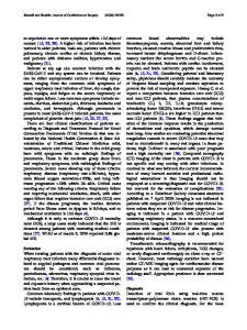
An 80-year-old Caucasian man presented for evaluation of systolic and diastolic heart failure. The patient’s medical history was notable for an IgG/kappa monoclonal protein identified 15 months prior by serum (SPEP) and urine protein electrophoresis studies (also had an elevated serum kappa to lambda free light chain ratio of 2.15) and deep vein thrombosis. Laboratory testing was notable for troponin I of 0.18 ng/mL, serum creatinine of 1.5 mg/dL, and blood urea nitrogen of 37 g/dL. An electrocardiogram (ECG) showed atrial fibrillation and left bundle branch block (LBBB) (Fig. 1), and angiography revealed only mild non-obstructive coronary artery disease. Echocardiogram revealed left ventricular ejection fraction of 40%, mild pulmonary hypertension, restrictive diastolic filing pattern, and moderate to severe left ventricular hypertrophy (Fig. 1). A technetium pyrophosphate scan was markedly abnormal and consistent with cardiac infiltrative disease in keeping with cardiac amyloidosis (Fig. 1). Based upon the patient’s clinical and radi
Data Loading...