Intraductal Hepatocellular Carcinoma without Parenchymal Tumor: A Case Report
- PDF / 267,913 Bytes
- 3 Pages / 595.276 x 790.866 pts Page_size
- 36 Downloads / 321 Views
CASE REPORT
Intraductal Hepatocellular Carcinoma without Parenchymal Tumor: A Case Report Banumathi Ramakrishna & Gautam Jitendra Shah & Frederick Vyas
Published online: 4 August 2011 # Springer Science+Business Media, LLC 2011
Abstract Introduction Obstructive jaundice due to hepatocellular carcinoma is rare. We present a case of hepatocellular carcinoma presenting as an intraductal tumor, which was clinically and radiologically diagnosed as cholangiocarcinoma. Clinical Presentation A 59-year-old male was admitted with recurrent episodes of jaundice. He was found to have a tumor in the right hepatic duct extending into intrahepatic ducts, which was clinically and radiologically diagnosed as cholangiocarcinoma. Results The patient underwent right hepatectomy with excision of the bile duct and left hepaticojejunostomy. Histological examination revealed an intraductal moderately differentiated hepatocellular carcinoma. The rest of the liver parenchyma showed features secondary to biliary obstruction but no tumor. Conclusion A case of hepatocellular carcinoma presenting as an intraductal tumor with obstructive jaundice and no evidence of parenchymal tumor is presented. Keywords Obstructive jaundice . Hepatocellular carcinoma . Intraductal tumor . Bile duct
Introduction Hepatocellular carcinoma (HCC) presenting as an intraductal tumor with obstructive jaundice and without parenchymal B. Ramakrishna (*) : G. J. Shah Department of Pathology, Christian Medical College, Vellore 632004 Tamil Nadu, India e-mail: [email protected] F. Vyas Department of Surgery, Christian Medical College, Vellore, India
tumor is uncommon. This type of tumor is also known as icteric-type HCC [1, 2]. We present a case of intraductal HCC in a 59-year-old male who was clinically and radiologically diagnosed as cholangiocarcinoma and underwent right hepatectomy and hepaticojejunostomy. Histological examination showed an intraductal HCC and no tumor in the liver parenchyma.
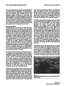
Case Report A 59-year-old male presented with recurrent episodes of jaundice for 10 months associated with upper abdominal pain, low-grade fever, and pruritus. There was no history of vomiting, hematemesis, melena, or altered bowel habits. He had loss of appetite and weight loss of 10 kg over this period. He was investigated elsewhere, and magnetic resonance cholangiopancreatogram done was suggestive of hilar cholangiocarcinoma. He was a diabetic and hypertensive on treatment. Investigations Liver function parameters showed increase in bilirubin and alkaline phosphatase and transaminitis. Viral markers were negative. CA19-9 was 245 U/ml. The serum alphafetoprotein level was not estimated. Ultrasound of the abdomen showed an echogenic heterogeneous mass in the hilum anterior to the portal vein with central intrahepatic biliary radicle dilatation (IHBRD). He underwent left percutaneous transhepatic biliary drain (PTBD) insertion. Combined magnetic resonance and computed tomography examination of the abdomen showed a heterogeneously enhancing mass measuring 30×24 mm at the h
Data Loading...