Ischemic chiasmal syndrome associated with posterior communicating artery (PCoA) and tuberothalamic artery (TA) infarcti
- PDF / 1,475,917 Bytes
- 4 Pages / 595.276 x 790.866 pts Page_size
- 97 Downloads / 249 Views
QUIZ CASES
Ischemic chiasmal syndrome associated with posterior communicating artery (PCoA) and tuberothalamic artery (TA) infarction: a case report Ceyla Ataç 1
&
Ayşın Kısabay Ak 2 & Melike Batum 2 & Semih Arı 2 & Gülgün Yılmaz Ovalı 3 & Neşe Çelebisoy 4
Received: 9 September 2020 / Accepted: 12 November 2020 # Fondazione Società Italiana di Neurologia 2020
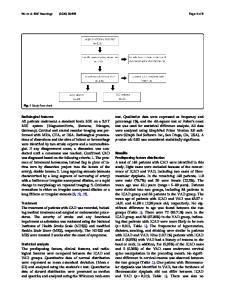
Abstract Lesions affecting the body of the optic chiasm typically produce bitemporal hemianopia. The blood supply comes from the anterior communicating artery, anterior cerebral, posterior communicating, posterior cerebral, and basilar arteries. We herein report a young patient admitted to the emergency department with acute confusion, left-sided hemiparesis, hemihypoesthesia, and dysarthria. Bitemporal hemianopia was detected after resolution of confusion. On cranial magnetic resonance imaging (MRI), infarction in the right anterolateral thalamus in the territory of tuberothalamic artery (TA) and in posterior chiasma in the territory of the posterior communicating artery (PCoA) was revealed. Cerebral MR angiography showed luminal irregularity of the PCoA. The patient was presented to draw attention to the rare entity ischemic chiasmal syndrome. Keywords Ischemic chiasmal syndrome . Posterior communicating artery . Tuberothalamic artery
Introduction The optic chiasm lies above the sphenoid bone, over the diaphragma sellae. Chiasmal disorders have been classified as intrinsic and extrinsic diseases: intrinsic causes including congenital malformations, trauma, chiasmal neuritis, and tumors arising within the chiasm and extrinsic etiologies resulting from mechanical compression due to sellar, parasellar, and cavernous sinus lesions which are more common. Pituitary tumors are known to be the most common cause of chiasmal syndrome in adults [1, 2]. Due to anatomical variations, chiasmal disorders can present with different visual symptoms and signs. Bitemporal
hemianopia is the classic finding of lesions affecting the body of the chiasm. The whole hemifield, upper or lower quadrants, can be affected. However, both due to individual variations and growth pattern of the compressive lesions, defects mainly involving the visual field of one eye can be seen when one optic nerve is more severely affected. Homonymous hemianopia can be detected in case of extension to the optic tractus [2]. Affection of the macular axons results in the loss of vision. Optic disc pallor indicates a more chronic process [1, 2]. The blood supply is variable as well. Branches coming from the anterior communicating artery (ACoA) and anterior cerebral artery (ACA) supply the superior part, sparing the
* Ceyla Ataç [email protected]
Neşe Çelebisoy [email protected]
Ayşın Kısabay Ak [email protected]
1
Department of Neurology, SBU İzmir Bozyaka Education and Research Hospital, 35360 İzmir, Turkey
Melike Batum [email protected]
2
Department of Neurology, Celal Bayar University, 45000 Manisa, Turkey
Semih Arı [email protected]
3
Department of Radiology, Celal
Data Loading...