Left ventricular dysfunction postsurgical patent ductus arteriosus ligation in children: predictor factors analysis
- PDF / 798,537 Bytes
- 6 Pages / 595.276 x 790.866 pts Page_size
- 30 Downloads / 338 Views
(2019) 14:168
RESEARCH ARTICLE
Open Access
Left ventricular dysfunction postsurgical patent ductus arteriosus ligation in children: predictor factors analysis Mohamed Abdel-Bary1* , Khaled Abdalla Abdel-Baseer2 , Ahmed Fathy Abdel-Latif3 , Mohamed Abdella Abdel-Naser4 , Mahmoud Nafie5 and Karam Mosallam Eisa1
Abstract Objective: To identify the predictor factors of left ventricular (LV) dysfunction following patent ductus arteriosus (PDA) surgical ligation. Background: PDA is viewed as a noticeable amongst the most widely recognized congenital heart defects in children and its closure is responsible for many hemodynamic changes that require intervention and care. Methods: A retrospective study included fifty children with isolated PDA treated by surgical ligation from June 2015 to June 2018. The LV dimensions and systolic function were assessed by two-dimensional echocardiography pre and post PDA ligation. All cases were followed-up on the first-day, 1 month and 6 months post ligation. Results: The mean age of cases was 15.78 ± 7.58 months and 72% were females. The mean duct size was 4.08 ± 1.25 mm. There was a marked decrease in LVEDd, LA/Ao, EF and FS in the first-day post ligation contrasted with pre ligation values. Moreover, an amazing decline in LVEDd and LA/Ao ratio was observed 1 month post ligation contrasted with the early post ligation status with asynchronous improvement of FS and EF at one and 6 months postoperatively. Conclusion: PDA ligation is associated with a noteworthy LV systolic dysfunction within the first day post ligation; that in a significant number of patients may require anti-failure measures, prolong the hospital stay and necessitate a regular follow up and monitoring of LV function. PDA size, age, preoperative LVEDd and FS can be considered as predictor factors for suspicion of acute decrease in the LV systolic function early post PDA ligation. Trial registration: ClinTrial.Gov NCT04018079. Keywords: PDA ligation, Congenital heart disease, LV systolic function and dimensions, LA/Ao ratio
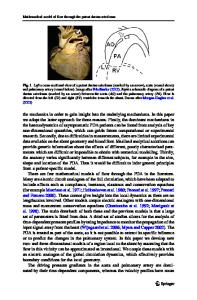
Background Ductus arteriosus is a shunt in the fetal circulation between the pulmonary artery and the proximal descending aorta [1]. A confined PDA is a noticeable amongst the most common congenital heart defects (CHD); as its incidence up to 8 for every 10,000 live births among term infants [2, 3]. The left to right shunt via a hemodynamically noteworthy PDA causes pulmonary over-flooding that result in the left ventricle (LV) volume over-burden and remodeling, and it compensates * Correspondence: [email protected] 1 Department of cardiothoracic surgery, Qena Faculty of Medicine, South Valley University, Safaga Road, Qena 83523, Egypt Full list of author information is available at the end of the article
by expanding stroke volume. Congestive heart failure (CHF) occurs in cases with greater shunts [4]. PDA may be associated with cardiac or respiratory morbidity. So, the closure should be performed once diagnosed. Surgical ligation indicated in huge PDA and not appropriate for percutane
Data Loading...