Locally Advanced Gastric Adenocarcinoma with Impressive Response to Hemostatic Radiation: the Possible Role of p53 Statu
- PDF / 4,444,913 Bytes
- 4 Pages / 595.276 x 790.866 pts Page_size
- 71 Downloads / 280 Views
CASE REPORT
Locally Advanced Gastric Adenocarcinoma with Impressive Response to Hemostatic Radiation: the Possible Role of p53 Status and Eosinophilic Infiltrate Abraão Ferreira Lopes Dornellas 1 & Marcus Fernando Kodama Pertille Ramos 1 & Marina Alessandra Pereira 1 & Leonardo Cardili 1 & Andre Roncon Dias 1 & Tiago Biachi de Castria 1,2 Accepted: 13 September 2020 # Springer Science+Business Media, LLC, part of Springer Nature 2020
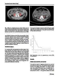
Case Presentation A 63-year-old female patient was referred to our hospital with symptoms of epigastric pain and weight loss for 5 months. Despite not being severe, vomiting was a frequent complain. Her past medical history was significant for hypertension, hypothyroidism, and dyslipidemia. Upper digestive endoscopy demonstrated an ulcerated lesion starting at the angular incisure, compromising circumferentially the antrum. The pylorus was infiltrated hardly allowing access to the duodenum (Fig. 1). Biopsy revealed a high-grade malignant epithelioid neoplasm showing solid architecture. The neoplastic cells were intermingled by a mixed inflammatory infiltrate rich in eosinophils and displayed high nuclear-cytoplasmic ratio with single or few punctate nucleoli. Diffuse immunolabelling for vimentin, high proliferative index (Ki67 > 80%), focal immunolabelling for pancytokeratin AE1/AE3, and CD56 were observed. CK7, CK20, CAM5.2, chromogranin A, S100 protein, and CD117 were negative. Helicobacter pylori was negative. Computed tomography (CT) scans showed a circumferential infiltrative lesion affecting the gastric antrum and measuring approximately 8.5 cm. Pancreatic invasion was suspected. Metastatic perigastric lymph nodes were also observed (the largest measured 4.5 cm) (Fig. 2). Laboratory exams such as hemoglobin, platelet, albumin, bilirubin, transaminases, and
* Abraão Ferreira Lopes Dornellas [email protected] 1
Cancer Institute, Hospital das Clinicas, University of Sao Paulo Medical School, Av Dr Arnaldo 251, São Paulo, SP 01249000, Brazil
2
Sirio-Libanes Hospital, Sao Paulo, Brazil
coagulogram were all within normal limits. The clinical stage was cT4b cN2 cM0 (IIIB). The patient underwent diagnostic laparoscopy to complement clinical staging. An extensive lesion with massive invasion in the pancreatic neck was noticed and a gastroenteric mechanical anastomosis performed due to gastric outlet obstruction and the poor clinical status at that time. Five days after the procedure the patient presented massive hematemesis, tumoral bleeding was confirmed and hemostatic radiotherapy indicated. The bleeding stopped after the total dose of 2000 cGy in 5 applications. Interestingly, a CT scan performed 8 weeks after radiation revealed an impressive radiologic response after the hemostatic radiotherapy (Fig. 2). The patient became asymptomatic and gained 20 kg with great improvement in her clinical status. Patient then underwent subtotal gastrectomy with D2 lymphadenectomy and Roux-en-Y reconstruction. Bursectomy was performed and the pancreatic capsule removed en bloc with the le
Data Loading...