Matrix metalloproteinase-9 expression in the Seoul-type keratoprosthesis implanted corneas with concurrent cultivated au
- PDF / 373,879 Bytes
- 4 Pages / 595.276 x 790.866 pts Page_size
- 63 Downloads / 245 Views
LETTER TO THE EDITOR
Matrix metalloproteinase-9 expression in the Seoul-type keratoprosthesis implanted corneas with concurrent cultivated autologous oral mucosal epithelial cell tranplantation Sang-Mok Lee & Mee-Kum Kim & Mi-Sun Shin & Won-Ryang Wee
Received: 17 November 2011 / Revised: 22 December 2011 / Accepted: 29 December 2011 / Published online: 18 January 2012 # Springer-Verlag 2012
Dear Editor, We evaluated the preliminary effect of cultivated autologous oral mucosal epithelial transplantation (COMET) in rabbit eyes with Seoul-type keratoprosthesis (SKPro), and found that COMET might suppress matrix metalloproteinase (MMP)-9 expressions. In keratoprosthetic eyes, the activities of gelatinolytic MMPs (MMP2 and 9) were reported to be increased postoperatively [1]. We were interested in the early stabilization of the
Presented in part at the ARVO, Ft Lauderdale, Florida, USA, May 2008.The authors have no relevant financial interest in this article. The authors have full control of all primary data, and they agree to allow Graefe's Archive for Clinical and Experimental Ophthalmology to review their data upon request. S.-M. Lee : M.-K. Kim (*) : W.-R. Wee Department of Ophthalmology, Seoul National University College of Medicine, 103 Daehak-ro, Jongno-gu, Seoul 110-799, Korea e-mail: [email protected] S.-M. Lee : M.-K. Kim : M.-S. Shin : W.-R. Wee Laboratory of Corneal Regenerative Medicine and Ocular Immunology, Artificial Eye Center, Clinical Research Institute Seoul National University Hospital, Seoul, Korea
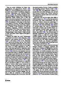
surgical wound after S-KPro implantation which had been developed to treat intractable ocular surface diseases [2–4], and thought that COMET, which was also introduced as a substitute for corneal epithelium, could be helpful in this aspect [5, 6]. The right corneas of six New Zealand white male rabbits were injured by 0.4 N NaOH to generate a limbal deficiency model (Fig. 1a). Their oral mucosas were harvested and the obtained cells in trypsin–EDTA solution were cultured on an human amniotic membrane (hAM) at an initial cell density of 1.0×10³cells/cm2 with mitomycin C-inactivated 3T3-J2 cells. After 2-weeks in submerged culture, they were air-lifted for another 2 weeks. More than 4 weeks after alkali burn injury (Fig. 1b), the S-KPro was implanted as mentioned previously [2, 7]. Thereafter, COMET was done and then temporary hAM was patched into the right eyes of three rabbits (COMET group, Fig. 1c). The remaining three rabbits underwent S-KPro implantation with hAM in their right eyes (control group). Postoperative management was treated as described previously [8]. Corneal new vessels and stromal melt were checked. The rabbits were sacrificed at 2, 3, and 8 weeks and evaluated with hematoxylin–eosin stain and immunohistochemical stain for CK4 and MMP-9. Mouse anti-CK4 antibody (VP-C399; Chemicon International, Temecula, CA, USA) 1:100 and mouse anti-MMP9 (MAB3309; Chemicon) 1:500
620
Graefes Arch Clin Exp Ophthalmol (2013) 251:619–622
Fig. 1 Anterior segment photos which show study methods and foll
Data Loading...