MRI brain tumor detection using optimal possibilistic fuzzy C-means clustering algorithm and adaptive k-nearest neighbor
- PDF / 3,292,989 Bytes
- 14 Pages / 595.276 x 790.866 pts Page_size
- 29 Downloads / 308 Views
ORIGINAL RESEARCH
MRI brain tumor detection using optimal possibilistic fuzzy C‑means clustering algorithm and adaptive k‑nearest neighbor classifier D. Maruthi Kumar1 · D. Satyanarayana2 · M. N. Giri Prasad1 Received: 6 March 2020 / Accepted: 29 July 2020 © Springer-Verlag GmbH Germany, part of Springer Nature 2020
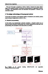
Abstract Brain tumor characterizes the aggregation of abnormal cells in specific tissues of the brain zone. The prior distinguishing proof of brain tumors has a huge influence on the treatment and recovery of the patient. The identification of a brain tumor and its evaluation is commonly a troublesome and tedious assignment. For effective classification and grading of brain tumor images, in this paper, we present an automatic MRI brain tumor classification system. The proposed work consists of four modules namely, pre-processing, feature extraction, classification, and segmentation. Initially, the noise present in the input image is removed using the Median Filter because the noises present in the input images will affect the accuracy of the classification process. At once, the images are converted into 3 × 3 blocks. Then, the texture features are extracted from the pre-processed image. After the feature extraction process, the features are given to the adaptive k-nearest neighbor classifier to classify an image as normal or abnormal. Later, the tumor regions are segmented with the help of the optimal possibilistic fuzzy C-means clustering algorithm. Both classification and the segmentation appearance technique are evaluated in terms of accuracy; sensitivity as well as specificity. For experimental analysis two dataset are utilized namely, BRATS MICCAI brain tumor dataset and publically available dataset. Keywords Median filter · AKNN · OPFCM
1 Introduction The brain is a complex organ of the human body and acts through billions of cells (Ljubimova et al 2017). Brain Tumors are classified based on the location of their origin and its malignancy (Dong et al 2017). A tumor is an abnormal development of brain tissues it can be dangerous or non-carcinogenic (Suhas and Venugopal 2017). As a result of the uncontrolled proliferation of tumors, tumors have abnormally growing tissues, and this tissue has no physiological function in the brain and causes increased volume and pressure in the brain, but also causes irregular nerve symptoms (Bahadure et al. 2017). A few disclosures, for example, X-rays, ultrasound, radiotherapy, MRI or computed tomography, and the development of devices that can create * D. Maruthi Kumar [email protected] 1
ECE, Jawaharlal Nehru Technological University, Anantapur, Ananthapuramu, Andhra Pradesh, India
ECE, Rajeev Gandhi Memorial College of Engineering and Technology, Nandyal, Andhra Pradesh, India
2
medical images have encouraged the advancement of the absolute most proficient investigation instruments in medication (Sharma et al., 2014). MRI is great therapeutic imaging, especially for brain imaging. The MRI classification strategies need diverse image highli
Data Loading...