Partially reversible confluent white matter lesions in a Caucasian child with moyamoya disease
- PDF / 2,948,782 Bytes
- 4 Pages / 595.276 x 790.866 pts Page_size
- 24 Downloads / 328 Views
LETTER TO THE EDITOR
Partially reversible confluent white matter lesions in a Caucasian child with moyamoya disease Ana Filipa Geraldo 1
&
Cátia Leitão 2 & Joana Nunes 1 & Marta Vila-Real 2
Received: 24 June 2020 / Accepted: 30 July 2020 # Springer-Verlag GmbH Germany, part of Springer Nature 2020
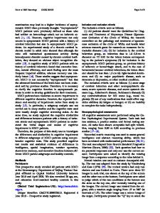
Dear Editor: We read with much interest a recent article published in Child’s Nervous System by Shi Jun et al. reporting the case of a 4-year-old girl with moyamoya disease (MMD) presenting white matter lesions on brain magnetic resonance imaging (MRI) that partially regressed over time with medical treatment alone [1]. We herein report a second pediatric patient with bilateral MMD-associated white matter abnormalities that markedly improved on a follow-up brain MRI before any surgical treatment was performed. This 5-year-old Caucasian boy was referred to our institution due to recurrent headaches. Physical and neurologic examinations were normal. Brain MRI was requested by the pediatric neurologist because the child had a 12-yearold brother with MMD diagnosed a few years earlier in the aftermath of an acute ischemic stroke. This older brother subsequently underwent neurosurgical revascularization, without new clinical events nor silent MRI lesions on follow-up. Initial brain MRI of the younger brother revealed striking bilateral, confluent hyperintense areas on T2-weighted and fluid-attenuated imaging recovery (FLAIR) images involving the peri-ventricular, deep, and subcortical supratentorial white matter, without associated mass effect. No other parenchymal abnormalities were depicted, namely signs of previous ischemic strokes or hemorrhagic infarcts
* Ana Filipa Geraldo [email protected] 1
Department of Medical Imaging, Diagnostic Neuroradiology Unit, CHVNG/E- Centro Hospitalar Vila Nova de Gaia/Espinho, Vila Nova de Gaia, Portugal
2
Department of Pediatrics, CHVNG/E- Centro Hospitalar Vila Nova de Gaia/Espinho, Vila Nova de Gaia, Portugal
(Fig. 1a–c). Magnetic resonance angiography with time-offlight (MRA-TOF) technique showed steno-occlusive changes of the vessels of the circle of Willis bilaterally as well as multiple, thin, tortuous lenticulostriate moyamoya vessels (Fig. 1d). Both arterial and white matter abnormalities were asymmetric, more prominent on the right side (Fig. 1a–c). Blood tests were unremarkable. As the imaging findings fulfilled the criteria of “definitive” MMD and the patient had a first-degree sibling affected with MMD, the diagnosis of familial MMD was assumed [2]. Interestingly, the older brother did not present white matter signal abnormalities at clinical presentation or at follow-up. The younger child was then referred to an outside neurosurgery department with a dedicated pediatric staff for further management. The best timing to perform revascularization surgery in this specific case presenting only with headaches is under evaluation and dependent on final acceptance by the parents. In the meanwhile, the child has been treated with an antiplatelet agent (aspirin, 3.8 mg
Data Loading...