Planar Laser-Induced Fluorescence Diagnostics of Pulsed Laser Ablation of Silicon
- PDF / 1,100,905 Bytes
- 6 Pages / 414.72 x 648 pts Page_size
- 59 Downloads / 293 Views
"**Combustion Research
Facility, Sandia National Laboratories, Livermore, CA
ABSTRACT Planar laser-induced fluorescence has been used to acquire time sequence images of ground-state, neutral Si and SiO during laser ablation of an Si target in vacuum and in the presence of a background gas at a fluence of 3-4 J/cm 2. The SiO images, taken in air, strongly suggest that the observed SiO is created through reaction of silicon with oxygen at the contact front as the plume expands. INTRODUCTION Pulsed laser deposition (PLD) is an attractive method to deposit a wide range of thin films [1]. The physics leading to plume formation is complex, resulting in a rich variety of plume phenomena, depending on target material, fluence, and the presence or absence of a background gas. Time-resolved imaging can shed light on some of the complex processes in the plumne. Several different methods have been employed to image PLD plumes, including emission, dye laser absorption, shadow photography, and planar laser-induced fluorescence (PLIF). (See Geohegan [21 for a review of these studies.) Planar laser-induced fluorescence is particularly useful, since it involves no line-of-sight averaging, is species-specific (like absorption) and can detect non-luminous ground state species. A number of groups have used single-point or one-dimensional laser-induced fluorescence for plume studies [3, 4, 5]. However, there are only two reports of two-dimensional planar LIF imaging of a laser-ablation plume [6, 7].
In the present paper, we present some results of a PLIF study of the ablation of an Si target into vacuum and air. We have acquired time sequence images of ground-state Si and, in the experiments with air, ground-state SiO. The images of SiO are, to our knowledge, the first reported of a reactive intermediate species generated during pulsed laser deposition. EXPERIMENTAL The experiments were carried out in a stainless steel vacuum cube with quartz windows. The experimental setup is shown schematically in Figure 1. The target was a silicon wafer and was rotated continuously during the experiments. The ablation laser was an injectionseeded 10 Hz excimer laser running on KrF with a nominal energy per pulse of 175 mJ. The excimer laser beam was focused onto the target at normal incidence using a single 500 mm focal length aberration-corrected doublet lens. The spot size on the target was approximately 2.5 mm diameter, resulting in a fluence of 3-4 J/cm2 . An excimer-pumped dye laser was used for the fluorescence excitation. Coumarin 480 dye was used for both the Si and SiO measurements, with a nominal energy per pulse of 1 mJ and a bandwidth of 0.15 cm- 1 . The dye laser beam was transformed into a collimated sheet nominally 1 cm wide using a telescope made from a -64 mm cylindrical lens and a +500 mm spherical lens. The laser sheet was brought to a soft waist of thickness 0.02 cm. 33
Mat. Res. Soc. Symp. Proc. Vol. 388 0 1995 Materials Research Society
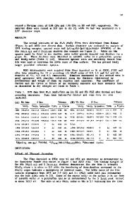
CCD camera7
filter
plume
rotating target (Si)
Figure 1: Schematic of experimental
Data Loading...