Radiation dose reduction considerations and imaging patterns of ground glass opacities in coronavirus: risk of over expo
- PDF / 989,560 Bytes
- 8 Pages / 595.276 x 790.866 pts Page_size
- 71 Downloads / 215 Views
CHEST RADIOLOGY
Radiation dose reduction considerations and imaging patterns of ground glass opacities in coronavirus: risk of over exposure in computed tomography Mohammad Ahmmad Rawashdeh1 · Charbel Saade2 Received: 28 April 2020 / Accepted: 23 August 2020 © Italian Society of Medical Radiology 2020
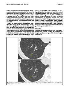
Abstract This article aims to summarize the available data on the severe acute respiratory syndrome coronavirus 2 (SAR-CoV-2) imaging patterns as well as reducing radiation dose exposure in chest computed tomography (CT) protocols. First, the general aspects of radiation dose in CT and radiation risk are discussed, followed by the effect of changing parameters on image quality. This article attempts to highlight some of the common chest CT signs that radiologists and emergency physicians are likely to encounter. With the increasing trend of using chest CT scans as an imaging tool to diagnose and monitor SARCoV-2, we emphasize that pattern recognition is the key, and this pictorial essay should serve as a guide to help establish correct diagnosis coupled with correct scanner parameters to reduce radiation dose without affecting imaging quality in this tragic pandemic the world is facing. Keywords COVID-19 · Multidetector computed tomography · Infections · Virology
Introduction The rapid rise of severe acute respiratory syndrome coronavirus 2 (SARS-CoV-2; previously known as 2019 novel coronavirus or 2019-nCoV) disease (COVID-19) has become a global emergency that has sent shockwaves in medicine to each corner of the globe [1]. The coronavirus infection took place in Wuhan, Hubei Province, China. In humans, coronaviruses are a series of viruses that cause the symptoms of upper respiratory infections which can become severe. The current status of mortality rates between China and Italy are similar with fatalities in mostly the elderly with known comorbidities [2]. Severe acute respiratory syndrome (SARS) and Middle East respiratory syndrome (MERS) have * Charbel Saade [email protected] Mohammad Ahmmad Rawashdeh [email protected] 1
Department of Allied Medical Sciences, Jordan University of Science and Technology, P.O.Box 3030, Irbid 22110, Jordan
Diagnostic Radiology Department, American University of Beirut Medical Center, P.O.Box 11‑0236, Riad El‑Solh, Beirut 1107 2020, Lebanon
2
mortality rates of 10% and 37%, in each of their respective continents [1, 3, 4]. This global emergency has resulted in increased utilization of chest imaging in the confirmation and progress of disease throughout their treatment. Over the last century, chest radiography was the first line of imaging in respiratory related pandemics that occurred such as Malaria, Influenza, Tuberculosis, SARS and HIV/ AIDS [5]. However, the explosion of computed tomography (CT) technology and the effect of lower doses in chest CT around the world have been heavily employed to monitor SARS-CoV-2 [6–10]. Standard chest CT doses can range from 3 to 4.8 mSv and low/ultra-low dose chest CT 0.13–1.5 mSv [11, 12]. A recent study demonstrated that n
Data Loading...