Renal Cell Carcinoma
Renal cell cancer (RCC) accounts for approximately 2–3 % of all human malignancies and shows a worldwide increase of incidence rate of 2 % per year [1]. It primarily affects men and women at the age of 50–70 years with a twofold higher incidence rate in m
- PDF / 526,839 Bytes
- 5 Pages / 439.37 x 666.14 pts Page_size
- 106 Downloads / 375 Views
39
Inga Peters, Maria Gabriel, Markus A. Kuczyk, and Axel S. Merseburger
39.1
General Facts
Renal cell cancer (RCC) accounts for approximately 2–3 % of all human malignancies and shows a worldwide increase of incidence rate of 2 % per year [1]. It primarily affects men and women at the age of 50–70 years with a twofold higher incidence rate in men. Clear cell carcinoma (ccRCC) is the most frequent histological subtype (~80–90 %) besides papillary (10–15 %) and chromophobe (4–5 %) subtype [2]. About 25–30 % of RCC patients present with a metastatic disease (mRCC) at the time of diagnosis. Patients with mRCC generally have a poor prognosis and the 5-year disease-specific survival for University of California Los Angeles integrated staging system (UISS) was 41 % in the low-risk group, 18 % in intermediate and 8 % in the high-risk group [3]. Aetiological risk factors for RCC seem to be smoking, hypertension and obesity [4]. The tumour node metastasis (TNM) staging system and the Fuhrman nuclear grade are commonly recommended for classification, diagnosis and prognosis of RCC patients (Table 39.1).
39.2
Symptoms, Classification and Grading
If a patient occurs with haematuria, flank pain and palpable abdominal mass, it is in all likelihood due to an already late stage renal cell cancer disease. Fortunately, nowadays about 50 % of renal masses are detected by using high-resolution imaging systems to investigate a range of non-specific symptoms. Paraneoplastic syndromes, which are summarised in Fig. 39.1, are frequently found (in about 30 %) to be associated with a RCC disease. Physical examination plays a minor role in diagnosing I. Peters • M. Gabriel • M.A. Kuczyk • A.S. Merseburger (*) Department of Urology and Urologic Oncology, Medizinische Hochschule Hannover (MHH), Carl-Neuberg-Str. 1, Hannover 30625, Germany e-mail: [email protected]; [email protected]; [email protected] A.S. Merseburger et al. (eds.), Urology at a Glance, DOI 10.1007/978-3-642-54859-8_39, © Springer-Verlag Berlin Heidelberg 2014
195
196
I. Peters et al.
Table 39.1 TNM classification of kidney tumours T – primary tumour T1a T1b T2a T2b T3a T3b T4 N – regional lymph nodes N1 N2 M – distant metastasis M1
Tumour ≤4 cm limited to the kidney Tumour >4 cm but not more than 7 cm Tumour >7 but not more that 10 cm Tumour >10 cm, limited to the kidney Tumour extends into renal vein or its segmental branches Tumour grossly extends into vena cava below diaphragm Gerota fascia invasion, adrenal gland invasion Metastasis in a single regional lymph node Metastasis in more than one regional lymph node Distant metastasis
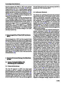
Fig. 39.1 Ultrasound image of a right kidney with suspicious lesion near the hilus
RCC, but in case of palpable abdominal mass, non-reducing varicocele, bilateral oedema of lower extremities or cervical lymphadenopathy, further radiological investigations should be urgently carried out. Generally, most renal tumours are diagnosed by abdominal ultrasound (US, see Fig. 39.1) or computer tomography (CT
Data Loading...