Renal granulomas mimicking as malignant renal masses on F18 FDG PET/CT in a case of urothelial carcinoma
- PDF / 5,263,692 Bytes
- 2 Pages / 595.276 x 790.866 pts Page_size
- 100 Downloads / 302 Views
IMAGE OF THE MONTH
Renal granulomas mimicking as malignant renal masses on F18 FDG PET/CT in a case of urothelial carcinoma Manikandan M V 1 & Archi Agrawal 1 & Santosh Menon 2 & Nilendu Purandare 1 & Sneha Shah 1 & Ameya Puranik 1 & Ganesh Bakshi 3 & Gagan Prakash 3 & Venkatesh Rangarajan 1 Received: 29 January 2020 / Accepted: 14 February 2020 # Springer-Verlag GmbH Germany, part of Springer Nature 2020
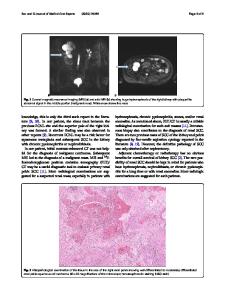
A 77-year-old male, case of high-grade papillary urothelial carcinoma, underwent trans urethral resection of bladder tumour (TURBT) followed by six weekly doses of intravesical bacillus Calmette-Guerin (BCG). During follow-up, urine cytology was positive for malignant cells. Cystoscopy and CT intravenous urogram did not reveal any bladder or ureteric lesions. 18F FDG PET/CECT was done and the maximal intensity projection image showed abnormal increased FDG uptake in the upper poles of both kidneys (image a). On coronal and axial images, increased FDG uptake noted in hypodense lesions involving upper poles of both kidneys (images b–j), measuring 22 mm on the right side, SUVmax of 12.92 (images g & e) and 14 mm on the left side, SUVmax of 10.02 (images j & h). With a suspicion of second primary malignancy or metastases, CT-guided biopsy of the renal
This article is part of the Topical Collection on Image of the month * Archi Agrawal [email protected] 1
Department of Nuclear Medicine and Molecular Imaging, Tata Memorial Hospital, Homi Bhabha National Institute, E. Borges Road, Parel, Mumbai 400 012, India
2
Department of Pathology, Tata Memorial Hospital, Homi Bhabha National Institute, Mumbai, India
3
Department of Uro Oncology, Tata Memorial Hospital, Homi Bhabha National Institute, Mumbai, India
lesion was done and histopathology revealed granulomatous inflammation with ill-formed granulomas with no evidence of malignancy. H&E-stained photomicrographs are depicted in image k (× 10) and image L (× 40) showing a non-necrotizing granuloma with epithelioid histiocytes admixed with few lymphocytes (L). The incidence of renal granuloma after intravesical instillation of BCG is very rare 0.1% [1]. It is more likely to be related to reflux of the intravesical BCG rather than hematogenous spread in post-TURBT patients [2]. They appear as FDG avid mass like lesions mimicking renal neoplasms and hence, proper histopathological evaluation is essential to avoid unnecessary surgical intervention. Other conditions which show false-positive FDG uptake in kidneys include granulomatosis with polyangiitis, infected renal cysts, focal pyelonephritis and oncocytomas [3–5].
Eur J Nucl Med Mol Imaging
Compliance with ethical standards Conflict of interest The authors declare that they have no conflict of interest. Informed consent Informed consent was taken from the patient for the publication of this case report and related images and histopathological data.
References 1.
Al-Qaoud T, Brimo F, Aprikian AG, Andonian S. BCG-related renal granulomas managed conservatively: a case series. Can Urol Assoc J. 2015;9(3–4):E200–3
Data Loading...