Robotic minimally invasive direct coronary artery bypass for Kawasaki disease
- PDF / 608,046 Bytes
- 3 Pages / 595.276 x 790.866 pts Page_size
- 95 Downloads / 379 Views
CASE REPORT
Robotic minimally invasive direct coronary artery bypass for Kawasaki disease Yorihiko Matsumoto1 · Satsuki Fukushima1 · Yusuke Shimahara1 · Naonori Kawamoto1 · Naoki Tadokoro1 · Soichiro Kitamura1 · Junjiro Kobayashi1 · Tomoyuki Fujita1 Received: 8 June 2019 / Accepted: 17 September 2019 © The Japanese Association for Thoracic Surgery 2019
Abstract Case 1: 17-year-old boy developed severe stenosis at the proximal site of the coronary aneurysm in the left anterior descending artery (LAD). Case 2: 16-year-old boy developed severe stenosis at the proximal site of the coronary aneurysm in the LAD. Case 3: 30-year-old woman developed severe stenosis of the distal portion of the coronary aneurysm in the LAD. Minimally invasive direct coronary artery bypass (MIDCAB) with robot-assisted left internal thoracic artery harvest was successfully performed without cardiopulmonary bypass in these three young patients with Kawasaki disease. This is the first case report of robot-assisted MIDCAB for Kawasaki disease. Keywords Kawasaki disease · Robotic-MIDCAB · Off-pump CABG
Introduction Kawasaki disease (KD) is acute systematic vasculitis of medium-sized arteries, mainly in children. KD causes aneurysmal formation in the proximal coronary arteries, typically in the proximal left anterior descending artery (LAD). The gold standard coronary artery bypass grafting (CABG) method for KD in children or adolescents is the left internal thoracic artery (ITA) to LAD graft, as this graft is durable and has growth potential [1, 2]. We report three cases of CABG via robot assisted mini-left thoracotomy for KD in adolescents and a young adult.
Case 1 A 17-year-old boy (height: 165 cm, bodyweight: 50 kg, body surface area: 1.52 m2) was diagnosed with KD at 9 years of age. Despite prompt treatment with aspirin and immunoglobulins, coronary angiography performed 2 months after diagnosis revealed an 11-mm-diameter aneurysm in the * Tomoyuki Fujita [email protected] 1
Department of Cardiovascular Surgery, National Cerebral and Cardiovascular Center, 5‑7‑1 Fujishirodai, Suita, Osaka 565‑8565, Japan
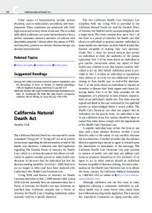
proximal LAD. Vitamin K antagonist (VKA) therapy was commenced, and there were no signs of myocardial ischemia from diagnosis to 17 years of age. At this time, enhanced computed tomography (CT) revealed that the aneurysm had increased to a diameter of 13 mm. Fluoroscopic coronary angiography showed 75% stenosis at the stem of the aneurysm in the proximal LAD (Fig. 1a), while exercise stress myocardial scintigraphy revealed a perfusion defect with redistribution at the anteroseptal region of the left ventricle. In the left semi-lateral decubitus position, bilateral arm ports of the da Vinci surgical system (Intuitive Surgical, Inc., Sunnyvale, CA) were inserted in the second and sixth intercostal spaces, and the camera port was inserted in the fourth intercostal space. The full length of the left ITA from the first to sixth intercostal spaces was dissected in a skeletonized fashion with monopolar spatula and bipolar forceps. Intra-
Data Loading...