Traumatic and degenerative cartilage lesions: arthroscopic differentiation using near-infrared spectroscopy (NIRS)
- PDF / 491,127 Bytes
- 6 Pages / 595.276 x 790.866 pts Page_size
- 3 Downloads / 245 Views
ARTHROSCOPY AND SPORTS MEDICINE
Traumatic and degenerative cartilage lesions: arthroscopic differentiation using near-infrared spectroscopy (NIRS) Gunter Spahn · Gernot Felmet · Gunther O. Hofmann
Received: 29 August 2012 / Published online: 1 May 2013 © European Union 2013
Abstract Introduction Cartilage lesions or defects are the most common finding during knee arthroscopy. During arthroscopy, it is often difficult to differentiate between degenerative and traumatic cartilage lesions. The study aimed to determine the impact of near-infrared spectroscopy (NIRS) on the distinction between traumatic and degenerative cartilage lesions in the medial femoral condyle (MFC). It was hypothesized that NIRS as able to distinguish between traumatic and degenerative cartilage lesions. Materials and methods Arthroscopic evaluation was performed in six patients who had undergone anterior cruciate ligament (ACL) reconstruction and in six patients who had undergone high tibial osteotomy (HTO). In both groups, a grade III cartilage lesion was present within the MFC. NIRS evaluation was performed with a special probe (arthrospec-one, Arthrospec GmbH, Jena, Germany). NIRS measurements produced semi-quantitative values ranging from 0 (heavily degenerated cartilage) to 100 (completely intact cartilage). Results The mean near-infrared-light absorption within the traumatic lesions in the MFC of the ACL group was 71.5 (range 61–80). In the HTO patients, this value was G. Spahn (*) Center of Trauma and Orthopaedic Surgery Eisenach, Sophienstr 16, 99817 Eisenach, Germany e-mail: [email protected] G. Felmet ARTICO Sports Clinic, Villingen-Schwenningen, Germany e-mail: [email protected] G. O. Hofmann Jena University Hospital and Trauma Center “Bergmannstrost”, Halle, Germany e-mail: [email protected]
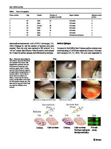
significantly (p 10,000 knee arthroscopies) performed the operations. In every patient, the joint surfaces were systematically evaluated at defined points. The patella was investigated at the medial, central, and lateral thirds (Patella_ medial, Patella_central and Patella_lateral, respectively). The same inspection was performed within the trochlea: medial, central (groove), and lateral (Trochlea_medial, Trochlea_central and Trochlea_lateral, respectively). Both the medial femoral condyle (MFC) and the lateral femoral condyle (LFC) were investigated within the mean bearing zone (mbz) as well as within the margin of the mean bearing zone. The same was done within the medial (TM) and lateral (LT) tibia. All regions of interest were evaluated using a special probe (arthrospec-one, Arthrospec GmbH, Jena, Germany), Fig. 3. The probe contains glass fibers. During the measurements, a NIR light is applied to the hyaline cartilage
13
1000
Arch Orthop Trauma Surg (2013) 133:997–1002
Table 2 Results of NIRS measurements within the chondral areas Mean
Patella_medial HTO ACL Patella_central HTO ACL Patella_lateral HTO ACL Trochlea_medial HTO ACL Trochlea_central HTO ACL Trochlea_lateral HTO ACL MFC_mbz H
Data Loading...