Radiation Therapy in the Management of Mantle Cell Lymphoma
Mantle cell lymphoma (MCL) comprises 3–6 % of non-Hodgkin lymphoma cases, with an annual incidence of 0.5 per 100,000 in Western countries. It is an aggressive malignancy that is usually disseminated at diagnosis; therefore, chemotherapy is typically the
- PDF / 784,678 Bytes
- 13 Pages / 504.57 x 720 pts Page_size
- 91 Downloads / 341 Views
10
Sarah A. Milgrom
Abstract
Mantle cell lymphoma (MCL) comprises 3–6 % of non-Hodgkin lymphoma cases, with an annual incidence of 0.5 per 100,000 in Western countries. It is an aggressive malignancy that is usually disseminated at diagnosis; therefore, chemotherapy is typically the backbone of treatment. However, MCL is exquisitely radiosensitive, and radiation therapy (RT) can be an important modality that contributes to cure in early-stage disease and provides durable palliation in advanced cases. In this chapter, patients with early- and advanced-stage MCL will be discussed, with a focus on the role of RT in management.
Background Mantle cell lymphoma (MCL) comprises 3–6 % of non-Hodgkin lymphoma cases, with an annual incidence of 0.5 per 100,000 in Western countries [1]. It is an aggressive malignancy that is usually disseminated at diagnosis; therefore, chemotherapy is typically the backbone of treatment. However, MCL is exquisitely radiosensitive, and radiation therapy (RT) can be an important modality that contributes to cure in early-stage disease and provides durable palliation in advanced cases. In this chapter, patients with
S.A. Milgrom, MD Department of Radiation Oncology, MD Anderson Cancer Center, 1515 Holcombe Blvd, Houston, TX 77030, USA e-mail: [email protected]
early- and advanced-stage MCL will be discussed, with a focus on the role of RT in management.
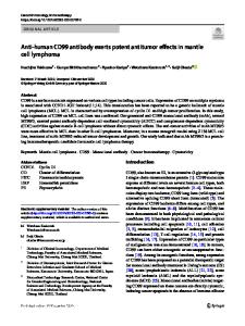
Early-Stage Mantle Cell Lymphoma Clinical Presentation, Early-Stage Case A healthy 69-year-old man was noted on a routine eye examination to have a pink growth extending along the inferior bulbar conjunctiva of the left eye (Fig. 10.1). The remaining ophthalmologic examination was normal. A biopsy of the conjunctival mass revealed a diagnosis of MCL. This patient is typical, with respect to demographics: the median age at diagnosis of MCL is 68 years, and the male-to-female ratio is 3:1 [1].
© Springer International Publishing Switzerland 2017 B.S. Dabaja, A.K. Ng (eds.), Radiation Therapy in Hematologic Malignancies, DOI 10.1007/978-3-319-42615-0_10
143
144
Fig. 10.1 MCL involving the inferior bulbar conjunctiva of the left eye
A careful history and physical examination was performed. The patient had an excellent performance status. He had no symptoms attributable to the eye. He denied constitutional symptoms of fever, night sweats, and weight loss. The conjunctival lesion was the only abnormality appreciated on physical examination. Special attention was paid to the nodal basins, liver, and spleen, all of which were unremarkable.
Pathology, Early-Stage Case An incisional biopsy of the conjunctival mass was performed. It should be noted that a fineneedle aspiration is typically insufficient to make the diagnosis of MCL. The biopsy revealed a glandular epithelium, largely replaced by lymphocytes with features typical of MCL, which arises from immature B-cells. The neoplastic cells tend to be small to medium sized, with irregular nuclear contours, inconspicuous nucleoli, and scant cytoplasm. This patient’s neoplasm was noted
Data Loading...