Reverse redistribution on 201 Tl SPECT in a patient with coronary artery ectasia
- PDF / 2,831,001 Bytes
- 4 Pages / 593.972 x 792 pts Page_size
- 75 Downloads / 280 Views
Department of Radiology, Faculty of Medicine, Kagawa University, Kita-gun, Kagawa, Japan
Received Aug 25, 2020; accepted Aug 25, 2020 doi:10.1007/s12350-020-02361-4
INTRODUCTION 201
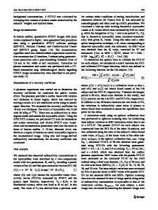
The reverse redistribution (RR) pattern on Tl single photon emission computed tomography (SPECT) may be defined as a normal or nearly normal perfusion in an early imaging (stress or rest acquisition after injection) and significantly smaller or less perfusion in a delayed rest imaging. We report the RR pattern observed on 201Tl SPECT due to an uncommon cause: coronary artery ectasia. CASE PRESENTATION A 61-year-old woman with atypical chest pain presented to our hospital. Transthoracic echocardiogram (TTE) showed hypokinesis from the base to mid inferior walls and apex, but her cardiac function was maintained. On rest early 201Tl SPECT, almost normal myocardial uptake was shown, although on rest delayed (3 hours) 201 Tl SPECT, a mild diffuse heterogeneous decreased uptake and a decreased marked focal uptake in the mid inferolateral wall were shown (Figure 1). The imaging findings were consistent with the RR pattern. Coronary artery angiography (CAG) and coronary computed
Reprint requests: Takashi Norikane, MD, PhD, Department of Radiology, Faculty of Medicine, Kagawa University, 1750-1 Ikenobe, Miki-cho, Kita-gun, Kagwa 761-0793, Japan; [email protected] J Nucl Cardiol 1071-3581/$34.00 Copyright Ó 2020 American Society of Nuclear Cardiology.
tomography angiography (CCTA) were performed for further evaluation. CAG and CCTA depicted a leftdominant coronary artery and both right and left dilated coronary arteries (Figure 2). On both CAG and CCTA images, contrast opacification was shown only up to the mid-vessel segment of the main coronary arteries, and contrast medium did not reach up to #3 or #15PD indicating slow blood flow because of excessive dilation. Hybrid 201Tl SPECT/CCTA images revealed that the area where the RR patten was seen was located in the distal left circumflex artery territory (Figure 3). Coronary artery ectasia (CAE) is defined as a diffuse dilatation with a diameter of more than 1.5 times normal coronary artery and is a comparably rare cardiovascular disorder. The clinical significance of CAE is not clear yet. Although most CAE patients are asymptomatic, some present with angina pectoris or acute coronary syndrome. The RR pattern is usually seen on an ischemic improvement region after reperfusion therapy for acute myocardial infarction. In addition, the RR pattern is also observed in chronic stable coronary artery disease, coronary artery spasm and other disorders including cardiac sarcoidosis and Chagas
Manabe et al Reverse redistribution on
Journal of Nuclear CardiologyÒ 201
Tl SPECT in a patient
Early image
Delayed image
Early image
Delayed image
Figure 1. Rest early and delayed (3 hours) 201Tl single photon emission computed tomography (SPECT) images aligned in standard short axis, vertical long axis, horizontal long axis, and polar map. The reverse redistribution pattern in the mid inferolateral wall
Data Loading...