Sonozaki syndrome in the spotlight of imaging
- PDF / 309,960 Bytes
- 2 Pages / 595.276 x 790.866 pts Page_size
- 42 Downloads / 303 Views
CLINICAL IMAGE
Sonozaki syndrome in the spotlight of imaging Susanna Guerrini 1
&
Nicola Giordano 2
&
Luca Volterrani 3
&
Bruno Frediani 4 & Maria Antonietta Mazzei 3
Received: 3 August 2020 / Revised: 30 September 2020 / Accepted: 18 October 2020 # International League of Associations for Rheumatology (ILAR) 2020
Presentation Sonozaki syndrome (SS) also known as pustulotic arthroosteitis (PAO) is a rare chronic inflammatory disease belonging to the group of spondyloarthritis (SpA), firstly described by Sonozaki [1]. Onset is frequently seen at age 30–40, with a similar prevalence between sexes. Sterile palmoplantar pustular lesions and anterior thoracic joint involvement (with 77% of manubrium sterni) are the most typical findings, although other joints can be involved (34% of axial, 32% of peripheral joints, and 13% of sacroiliitis). We present the case of a 30-year-old male with palmoplantar pustulosis (PPP) and painful swelling of the sternum, clavicles, and lower ribs. The patient had no family history of other rheumatic disorders. General physical examination resulted negative for clinical involvement of the spinal column, and of the sacroiliac and peripheral joints, with no signs or symptoms of infective disease. Laboratory findings showed mild increase of erythrocyte sedimentation rate (ESR) (50 mm/h) and C-reactive protein (CRP) (2.5 mg/dl, n.v. 0.5); other biochemical tests (blood count, serum electrolytes, creatinine, urea, uric acid, glucose, bilirubin, transaminases, creatine phosphokinase, LDH, alcaline phosphatase), protein levels with electrophoresis, and general tests of urine, immunoglobulins, and complement and anti-CCP were normal; rheumatoid factor (RA test and Waaler rose) and antinuclear antibodies (ANA) were negative. HLA typing resulted negative for aplotipo HLAB51, 52, and 27. Skin biopsy confirmed the suspected PAO
* Susanna Guerrini [email protected] 1
Unit of Diagnostic Imaging, Department of Radiological Sciences, Azienda Ospedaliero-Universitaria Senese, “Santa Maria alle Scotte” General Hospital, Viale Mario Bracci, 16, 53100 Siena, Tuscany, Italy
diagnosis. The evaluation of the patient at 6 and 12 months confirmed the complete recovery after therapy with 16 mg of metilprednisolone with descending sequences of dose for 2 months.
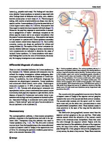
Discussion Sternocostoclavicular hyperostosis (SSCH) represented by diffuse sclerosis in the sternum, clavicle, or ribs in the advanced phase and by inflammatory enthesopathy of the costoclavicular ligament, small hyperostotic foci at the sternal end of ribs, and isolated periosteal reaction involving the clavicles in the early phase are characteristic radiological findings of SS, usually accompanied by erosive and sclerotic processes of the manubriosternal joint, as expression of arthritis [2] (Fig. 1). Spondylodiscitis in the vertebral involvement and sclerosis/hyperostosis in the sacroiliac joints may be also observed, whereas the peripheral joint involvement of SS is usually not severe and non-erosive. The latter may suggest the differen
Data Loading...