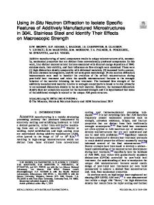
Specific features of two diffraction schemes for a widely divergent X-ray beam
- PDF / 646,550 Bytes
- 4 Pages / 612 x 792 pts (letter) Page_size
- 101 Downloads / 301 Views
RACTION AND SCATTERING OF IONIZING RADIATIONS
Specific Features of Two Diffraction Schemes for a Widely Divergent X-Ray Beam K. T. Avetyan, L. V. Levonyan, H. S. Semerjian, M. M. Arakelyan, and O. M. Badalyan Yerevan State University, Yerevan, 0025 Armenia e-mail: [email protected] Received April 28, 2014
Abstract—We investigated the specific features of two diffraction schemes for a widely divergent X-ray beam that use a circular diaphragm 30–50 μm in diameter as a point source of characteristic radiation. In one of the schemes, the diaphragm was set in front of the crystal (the diaphragm–crystal (d–c) scheme); in the other, it was installed behind the crystal (the crystal–diaphragm (c–d) scheme). It was established that the diffraction image in the c–d scheme is a topographic map of the investigated crystal area. In the d–c scheme at L = 2l (l and L are the distances between the crystal and the diaphragm and between the photographic plate and the diaphragm, respectively), the branches of hyperbolas formed in this family of planes (hkl) by the characteristic Kα and Kβ radiations, including higher order reflections, converge into one straight line. It is experimentally demonstrated that this convergence is very sensitive to structural inhomogeneities in the crystal under study. DOI: 10.1134/S1063774515010022
INTRODUCTION The diffraction pattern formed by a widely divergent beam (WDB) of characteristic X rays contains much information on the specific features of the crystal structure of an object under study, since many Bragg reflections are simultaneously detected in this case. Currently, there are two methods of X-ray WDB diffraction. In the Kossel method, a point X-ray source is located on the surface of the investigated object or under it; in the pseudo-Kossel method, the point source is located above the surface of an object studied [1–3]. These methods differ in how characteristic radiation is excited, i.e., forming a point radiation source. In this study we used two somewhat different schemes of WDB diffraction with a standard radiation source, i.e., an X-ray tube with a linear focal spot [4, 5]. In one scheme an X-ray beam passes through a diaphragm in the form of a funnel aperture (30–50 μm in diameter) in a tantalum plate and falls on a crystal under study. The diaphragm, crystal, and photographic plate are placed in a small chamber rotating around the diaphragm axis during exposure. It is only diffracted radiation that falls on a photographic plate mounted behind the crystal. The primary (nondiffracted) radiation is blocked by a trap, the position of which with respect to the radiation source does not change during chamber rotation [4]. In this configuration, referred to as the diaphragm–crystal (d–c)
scheme (Fig. 1a), the diaphragm works as a point source. When the diaphragm is installed close to the object studied, in front of it or behind it, the scheme is analogous to the Kossel scheme and the diffraction image does not differ from classical Kossel pattern. When the diaphragm is spaced by a distance
Data Loading...