Spinal cord subependymoma mimicking syringomyelia in a child: a case report
- PDF / 1,067,279 Bytes
- 5 Pages / 595.276 x 790.866 pts Page_size
- 27 Downloads / 319 Views
CASE REPORT
Spinal cord subependymoma mimicking syringomyelia in a child: a case report Masahiro Oishi 1,2
&
Hironori Fujisawa 1 & Katsuhiro Tsuchiya 1 & Yoshio Nakashima 1
Received: 15 September 2020 / Accepted: 20 October 2020 # Springer-Verlag GmbH Germany, part of Springer Nature 2020
Abstract Spinal cord subependymomas (SCSEs) in children are extremely rare, and no reports distinguishing SCSEs from syringomyelia have been published. We report a case of a 10-year-old boy who presented with torticollis, scoliosis, as well as pain that had begun in the posterior portion of the neck and progressed to the right shoulder and upper arm. Magnetic resonance imaging showed an intramedullary cyst-like lesion with the same signal intensity as that of cerebrospinal fluid. Idiopathic syringomyelia with scoliosis was first suspected, and a syrinx-subarachnoid space shunt was performed. After surgery, the lesion was slightly smaller; however, 2 years after surgery, it had re-grown, causing excruciating pain but no other symptoms. A second surgery was performed, and gross total resection was achieved. Pathological evaluation revealed SCSE. SCSE needs to be considered as a differential diagnosis for spinal centric cyst-like lesions in children. Keywords Spinal cord subependymoma . Syringomyelia . Child . Pediatric neurosurgery . Pediatric benign tumor . Scoliosis
Introduction
Case description
Subependymomas are slow-growing, World Health Organization grade I tumors [9] typically found in the fourth and lateral ventricles [6]. Spinal cord subependymomas (SCSEs) are rare, accounting for only 2% of all symptomatic spinal cord tumors. SCSEs usually occur in middle- and oldage individuals [2, 15, 18] and are extremely rare in children [15]. Owing to similar magnetic resonance imaging (MRI) characteristics, SCSEs may be difficult to differentiate from other spinal tumors, particularly ependymomas and astrocytomas [15, 18, 19]. SCSEs may be associated with syringomyelia, but differentiation between them is usually unnecessary. We report a case of SCSE in a 10-year-old child presenting with MRI findings similar to syringomyelia with scoliosis.
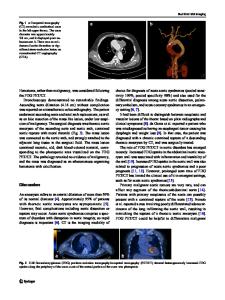
A 10-year-old boy presented with a 3-week history of progressive pain, initially in the posterior portion of the neck and subsequently in the right shoulder and upper arm. On admission, his right fourth and fifth fingers were numb and torticollis was evident; his McCormick classification (MMC) was grade 2. Neurological examination revealed no motor weakness, sensory loss, urinary retention, or defecation problems, and tendon reflexes were normal. X-rays revealed scoliosis (Fig. 1a). MRI showed an intramedullary central lesion extending from C5 to T2, which was hypointense on T1-weighted images and hyperintense on T2-weighted images, similar to cerebrospinal fluid (CSF) (Fig. 1b–e). There was no contrast enhancement (Fig. 1f, g), perilesional edema, Chiari malformation type I (CM-1) (Fig. 1d), or tethered spinal cord syndrome (Fig. 1h, i). Idiopathic syringomyelia with scoliosis rather th
Data Loading...